
Page 603 - Textbook of Pathology, 6th Edition
P. 603
587
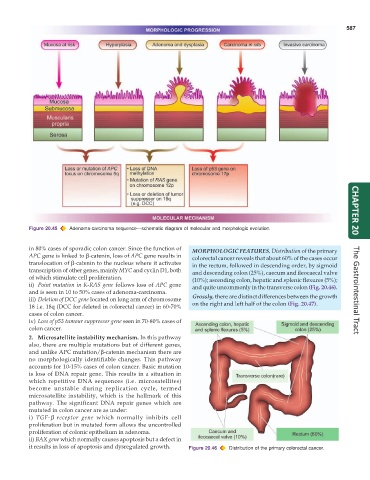
Figure 20.45 Adenoma-carcinoma sequence—schematic diagram of molecular and morphologic evolution. CHAPTER 20
in 80% cases of sporadic colon cancer. Since the function of MORPHOLOGIC FEATURES. Distribution of the primary
APC gene is linked to β-catenin, loss of APC gene results in colorectal cancer reveals that about 60% of the cases occur
translocation of β-catenin to the nucleus where it activates in the rectum, followed in descending order, by sigmoid
transcription of other genes, mainly MYC and cyclin D1, both and descending colon (25%), caecum and ileocaecal valve
of which stimulate cell proliferation. (10%); ascending colon, hepatic and splenic flexures (5%);
ii) Point mutation in K-RAS gene follows loss of APC gene and quite uncommonly in the transverse colon (Fig. 20.46).
and is seen in 10 to 50% cases of adenoma-carcinoma. The Gastrointestinal Tract
iii) Deletion of DCC gene located on long arm of chromosome Grossly, there are distinct differences between the growth
18 i.e. 18q (DCC for deleted in colorectal cancer) in 60-70% on the right and left half of the colon (Fig. 20.47).
cases of colon cancer.
iv) Loss of p53 tumour suppressor gene seen in 70-80% cases of
colon cancer.
2. Microsatellite instability mechanism. In this pathway
also, there are multiple mutations but of different genes,
and unlike APC mutation/β-catenin mechanism there are
no morphologically identifiable changes. This pathway
accounts for 10-15% cases of colon cancer. Basic mutation
is loss of DNA repair gene. This results in a situation in
which repetitive DNA sequences (i.e. microsatellites)
become unstable during replication cycle, termed
microsatellite instability, which is the hallmark of this
pathway. The significant DNA repair genes which are
mutated in colon cancer are as under:
i) TGF-β receptor gene which normally inhibits cell
proliferation but in mutated form allows the uncontrolled
proliferation of colonic epithelium in adenoma.
ii) BAX gene which normally causes apoptosis but a defect in
it results in loss of apoptosis and dysregulated growth. Figure 20.46 Distribution of the primary colorectal cancer.