
Page 606 - Textbook of Pathology, 6th Edition
P. 606
590
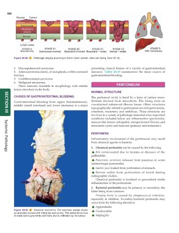
Figure 20.49 Pathologic staging according to Astler-Coller system. (Also see facing Table 20.12).
3. Mucoepidermoid carcinoma presenting clinical feature of a variety of gastrointestinal
4. Adenocarcinoma (rectal, of anal glands, within anorectal diseases. Table 20.13 summarises the main causes of
fistulas) gastrointestinal bleeding.
5. Undifferentiated carcinoma
6. Malignant melanoma.
These tumours resemble in morphology with similar PERITONEUM
lesion elsewhere in the body.
NORMAL STRUCTURE
CAUSES OF GASTROINTESTINAL BLEEDING The peritoneal cavity is lined by a layer of surface meso-
Gastrointestinal bleeding from upper (haematemesis), thelium derived from mesoderm. The lining rests on
middle (small intestinal) and lower (melaena) is a major vascularised subserosal fibrous tissue. Other structures
topographically related to peritoneum are retroperitoneum,
omentum, mesentery and umbilicus. These structures are
SECTION III
involved in a variety of pathologic states but a few important
conditions included below are inflammation (peritonitis),
tumour-like lesions (idiopathic retroperitoneal fibrosis and
mesenteric cysts) and tumours (primary and metastatic).
PERITONITIS
Inflammatory involvement of the peritoneum may result
from chemical agents or bacteria.
1. Chemical peritonitis can be caused by the following:
Bile extravasated due to trauma or diseases of the
Systemic Pathology
gallbladder.
Pancreatic secretions released from pancreas in acute
haemorrhagic pancreatitis.
Gastric juice leaked from perforation of stomach.
Barium sulfate from perforation of bowel during
radiographic studies.
Chemical peritonitis is localised or generalised sterile
inflammation of the peritoneum.
2. Bacterial peritonitis may be primary or secondary; the
latter being more common.
Primary form is caused by streptococcal infection,
especially in children. Secondary bacterial peritonitis may
occur from the following disorders:
Appendicitis
Figure 20.50 Anorectal carcinoma. The anorectal margin shows Cholecystitis
an ulcerated mucosa with thickened wall (arrow). The sectioned surface
of rectal wall is grey-white and fleshy due to infiltration by the tumour. Salpingitis