
Page 608 - Textbook of Pathology, 6th Edition
P. 608
592
The Liver, Biliary Tract and
Chapter 21
Chapter 21
Exocrine Pancreas
LIVER
NORMAL STRUCTURE
ANATOMY. The liver is the largest organ in the body
weighing 1400-1600 gm in the males and 1200-1400 gm in
the females. There are 2 main anatomical lobes—right and
left, the right being about six times the size of the left lobe.
The right lobe has quadrate lobe on its inferior surface and a
caudate lobe on the posterior surface. The right and left lobes
are separated anteriorly by a fold of peritoneum called the
falciform ligament, inferiorly by the fissure for the ligamentum
teres, and posteriorly by the fissure for the ligamentum
venosum (Fig. 21.1).
The porta hepatis is the region on the inferior surface of
the right lobe where blood vessels, lymphatics and common
hepatic duct form the hilum of the liver. A firm smooth layer
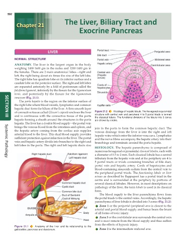
of connective tissue called Glisson’s capsule encloses the liver Figure 21.2 Histology of hepatic lobule. The hexagonal or pyramidal
and is continuous with the connective tissue of the porta structure with central vein and peripheral 4 to 5 portal triads is termed
the classical lobule. The functional divisions of the lobule into 3 zones
SECTION III
hepatis forming a sheath around the structures in the porta are shown by circles.
hepatis. The liver has a double blood supply—the portal vein
brings the venous blood from the intestines and spleen, and join in the porta to form the common hepatic duct. The
the hepatic artery coming from the coeliac axis supplies venous drainage from the liver is into the right and left
arterial blood to the liver. This dual blood supply provides hepatic veins which enter the inferior vena cava. Lymphatics
sufficient protection against infarction in the liver. The portal and the nerve fibres accompany the hepatic artery into their
vein and hepatic artery divide into branches to the right and branchings and terminate around the porta hepatis.
left lobes in the porta. The right and left hepatic ducts also
HISTOLOGY. The hepatic parenchyma is composed of
numerous hexagonal or pyramidal classical lobules, each with
a diameter of 0.5 to 2 mm. Each classical lobule has a central
tributary from the hepatic vein and at the periphery are 4 to
Systemic Pathology
5 portal tracts or triads containing branches of bile duct,
portal vein and hepatic artery. Cords of hepatocytes and
blood-containing sinusoids radiate from the central vein to
the peripheral portal triads. The functioning lobule or liver
acinus as described by Rappaport has a portal triad in the
centre and is surrounded at the periphery by portions of
several classical lobules. However, in most descriptions on
pathology of the liver, the term lobule is used in its classical
form.
The blood supply to the liver parenchyma flows from
the portal triads to the central veins. Accordingly, the hepatic
parenchyma of liver lobule is divided into 3 zones (Fig. 21.2):
Zone 1 or the periportal (peripheral) area is closest to the
arterial and portal blood supply and hence bears the brunt
of all forms of toxic injury.
Zone 3 or the centrilobular area surrounds the central vein
and is most remote from the blood supply and thus suffers
from the effects of hypoxic injury.
Figure 21.1 Anatomy of the liver and its relationship to the
gallbladder, pancreas and duodenum. Zone 2 is the intermediate midzonal area.