
Page 615 - Textbook of Pathology, 6th Edition
P. 615
599
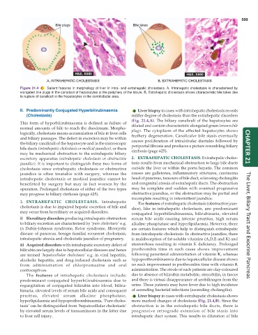
Figure 21.4 Salient features in morphology of liver in intra- and extrahepatic cholestasis. A, Intrahepatic cholestasis is characterised by
elongated bile plugs in the canaliculi of hepatocytes at the periphery of the lobule. B, Extrahepatic cholestasis shows characteristic bile lakes due
to rupture of canaliculi in the hepatocytes in the centrilobular area.
II. Predominantly Conjugated Hyperbilirubinaemia Liver biopsy in cases with intrahepatic cholestasis reveals
(Cholestasis) milder degree of cholestasis than the extrahepatic disorders
(Fig. 21.4,A). The biliary canaliculi of the hepatocytes are
This form of hyperbilirubinaemia is defined as failure of
normal amounts of bile to reach the duodenum. Morpho- dilated and contain characteristic elongated green-brown bile
plugs. The cytoplasm of the affected hepatocytes shows
logically, cholestasis means accumulation of bile in liver cells feathery degeneration. Canalicular bile stasis eventually
and biliary passages. The defect in excretion may be within causes proliferation of intralobular ductules followed by
the biliary canaliculi of the hepatocyte and in the microscopic periportal fibrosis and produces a picture resembling biliary
bile ducts (intrahepatic cholestasis or medical jaundice), or there cirrhosis (page 625). CHAPTER 21
may be mechanical obstruction to the extrahepatic biliary
excretory apparatus (extrahepatic cholestasis or obstructive 2. EXTRAHEPATIC CHOLESTASIS. Extrahepatic choles-
jaundice). It is important to distinguish these two forms of tasis results from mechanical obstruction to large bile ducts
cholestasis since extrahepatic cholestasis or obstructive outside the liver or within the porta hepatis. The common
jaundice is often treatable with surgery, whereas the causes are gallstones, inflammatory strictures, carcinoma
intrahepatic cholestasis or medical jaundice cannot be head of pancreas, tumours of bile duct, sclerosing cholangitis
benefitted by surgery but may in fact worsen by the and congenital atresia of extrahepatic ducts. The obstruction
operation. Prolonged cholestasis of either of the two types may be complete and sudden with eventual progressive
may progress to biliary cirrhosis (page 625). obstructive jaundice, or the obstruction may be partial and
incomplete resulting in intermittent jaundice.
1. INTRAHEPATIC CHOLESTASIS. Intrahepatic The features of extrahepatic cholestasis (obstructive jaun-
cholestasis is due to impaired hepatic excretion of bile and dice), like in intrahepatic cholestasis, are: predominant
may occur from hereditary or acquired disorders. conjugated hyperbilirubinaemia, bilirubinuria, elevated
i) Hereditary disorders producing intrahepatic obstruction serum bile acids causing intense pruritus, high serum
to biliary excretion are characterised by ‘pure cholestasis’ e.g. alkaline phosphatase and hyperlipidaemia. However, there
in Dubin-Johnson syndrome, Rotor syndrome, fibrocystic are certain features which help to distinguish extrahepatic The Liver, Biliary Tract and Exocrine Pancreas
disease of pancreas, benign familial recurrent cholestasis, from intrahepatic cholestasis. In obstructive jaundice, there
intrahepatic atresia and cholestatic jaundice of pregnancy. is malabsorption of fat-soluble vitamins (A,D,E and K) and
ii) Acquired disorders with intrahepatic excretory defect of steatorrhoea resulting in vitamin K deficiency. Prolonged
bilirubin are largely due to hepatocellular diseases and hence prothrombin time in such cases shows improvement
are termed ‘hepatocellular cholestasis’ e.g. in viral hepatitis, following parenteral administration of vitamin K, whereas
alcoholic hepatitis, and drug-induced cholestasis such as hypoprothrombinaemia due to hepatocellular disease shows
from administration of chlorpromazine and oral no such improvement in prothrombin time with vitamin K
contraceptives. administration. The stools of such patients are clay-coloured
The features of intrahepatic cholestasis include: due to absence of bilirubin metabolite, stercobilin, in faeces
predominant conjugated hyperbilirubinaemia due to and there is virtual disappearance of urobilinogen from the
regurgitation of conjugated bilirubin into blood, biliru- urine. These patients may have fever due to high incidence
binuria, elevated levels of serum bile acids and consequent of ascending bacterial infections (ascending cholangitis).
pruritus, elevated serum alkaline phosphatase, Liver biopsy in cases with extrahepatic cholestasis shows
hyperlipidaemia and hypoprothrombinaemia. ‘Pure choles- more marked changes of cholestasis (Fig. 21.4,B). Since the
tasis’ can be distinguished from ‘hepatocellular cholestasis’ obstruction is in the extrahepatic bile ducts, there is
by elevated serum levels of transaminases in the latter due progressive retrograde extension of bile stasis into
to liver cell injury. intrahepatic duct system. This results in dilatation of bile