
Page 621 - Textbook of Pathology, 6th Edition
P. 621
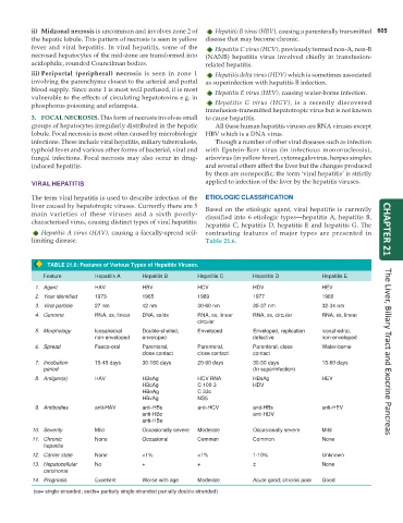
ii) Midzonal necrosis is uncommon and involves zone 2 of Hepatitis B virus (HBV), causing a parenterally transmitted 605
the hepatic lobule. This pattern of necrosis is seen in yellow disease that may become chronic.
fever and viral hepatitis. In viral hepatitis, some of the Hepatitis C virus (HCV), previously termed non-A, non-B
necrosed hepatocytes of the mid-zone are transformed into (NANB) hepatitis virus involved chiefly in transfusion-
acidophilic, rounded Councilman bodies. related hepatitis.
iii) Periportal (peripheral) necrosis is seen in zone 1 Hepatitis delta virus (HDV) which is sometimes associated
involving the parenchyma closest to the arterial and portal as superinfection with hepatitis B infection.
blood supply. Since zone 1 is most well perfused, it is most Hepatitis E virus (HEV), causing water-borne infection.
vulnerable to the effects of circulating hepatotoxins e.g. in
phosphorus poisoning and eclampsia. Hepatitis G virus (HGV), is a recently discovered
transfusion-transmitted hepatotropic virus but is not known
3. FOCAL NECROSIS. This form of necrosis involves small to cause hepatitis.
groups of hepatocytes irregularly distributed in the hepatic All these human hepatitis viruses are RNA viruses except
lobule. Focal necrosis is most often caused by microbiologic HBV which is a DNA virus.
infections. These include viral hepatitis, miliary tuberculosis, Though a number of other viral diseases such as infection
typhoid fever and various other forms of bacterial, viral and with Epstein-Barr virus (in infectious mononucleosis),
fungal infections. Focal necrosis may also occur in drug- arbovirus (in yellow fever), cytomegalovirus, herpes simplex
induced hepatitis. and several others affect the liver but the changes produced
by them are nonspecific; the term ‘viral hepatitis’ is strictly
VIRAL HEPATITIS applied to infection of the liver by the hepatitis viruses.
The term viral hepatitis is used to describe infection of the ETIOLOGIC CLASSIFICATION
liver caused by hepatotropic viruses. Currently there are 5 Based on the etiologic agent, viral hepatitis is currently
main varieties of these viruses and a sixth poorly- classified into 6 etiologic types—hepatitis A, hepatitis B,
characterised virus, causing distinct types of viral hepatitis:
hepatitis C, hepatitis D, hepatitis E and hepatitis G. The
Hepatitis A virus (HAV), causing a faecally-spread self- contrasting features of major types are presented in CHAPTER 21
limiting disease. Table 21.6.
TABLE 21.6: Features of Various Types of Hepatitis Viruses.
Feature Hepatitis A Hepatitis B Hepatitis C Hepatitis D Hepatitis E
1. Agent HAV HBV HCV HDV HEV
2. Year identified 1973 1965 1989 1977 1980
3. Viral particle 27 nm 42 nm 30-60 nm 35-37 nm 32-34 nm
4. Genome RNA, ss, linear DNA, ss/ds RNA, ss, linear RNA, ss, circular RNA, ss, linear
circular
5. Morphology Icosahedral Double-shelled, Enveloped Enveloped, replication Icosahedral,
non-enveloped enveloped defective non-enveloped
6. Spread Faeco-oral Parenteral, Parenteral, Parenteral, close Water-borne
close contact close contact contact The Liver, Biliary Tract and Exocrine Pancreas
7. Incubation 15-45 days 30-180 days 20-90 days 30-50 days 15-60 days
period (In superinfection)
8. Antigen(s) HAV HBsAg HCV RNA HBsAg HEV
HBcAg C 100-3 HDV
HBeAg C 33c
HBxAg NS5
9. Antibodies anti-HAV anti-HBs anti-HCV anti-HBs anti-HEV
anti-HBc anti-HDV
anti-HBe
10. Severity Mild Occasionally severe Moderate Occasionally severe Mild
11. Chronic None Occasional Common Common None
hepatitis
12. Carrier state None <1% <1% 1-10% Unknown
13. Hepatocellular No + + ± None
carcinoma
14. Prognosis Excellent Worse with age Moderate Acute good; chronic poor Good
(ss= single-stranded; ss/ds= partially single-stranded partially double-stranded)