
Page 624 - Textbook of Pathology, 6th Edition
P. 624
608
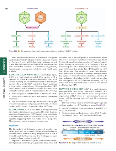
Figure 21.9 Consequences of coinfection versus superinfection in combined HDV-HBV infection.
HDV infection is worldwide in distribution though the accidental cuts and needle-pricks in health workers. About
incidence may vary in different countries. Endemic regions 90% of post-transfusion hepatitis is of hepatitis C type. About
are Southern Europe, Middle-East, South India and parts of 1-2% of volunteer blood donors and up to 5% of professional
Africa. The high-risk individuals for HDV infection are the blood donors are carriers of HCV. Hepatitis C has an
same as for HBV infection i.e. intravenous drug abusers, incubation period of 20-90 days (mean 50 days). Clinically,
homosexuals, transfusion recipients, and health care acute HCV hepatitis is milder than HBV hepatitis but HCV
workers. has a higher rate of progression to chronic hepatitis than
HBV. Persistence of infection and chronic hepatitis are the
SECTION III
HEPATITIS DELTA VIRUS (HDV). The etiologic agent, key features of HCV. Occurrence of cirrhosis after 5 to 10
HDV, is a small single-stranded RNA particle with a years and progression to hepatocellular carcinoma are other
diameter of 36 nm. It is double-shelled—the outer shell late consequences of HCV infection. Currently, HCV is
consists of HBsAg and the inner shell consists of delta antigen considered more important cause of chronic liver disease
provided by a circular RNA strand. It is highly infectious worldwide than HBV.
and can induce hepatitis in any HBsAg-positive host. HDV
replication and proliferation takes place within the nuclei of HEPATITIS C VIRUS (HCV). HCV is a single-stranded,
liver cells. Markers for HDV infection include the following: enveloped RNA virus, having a diameter of 30-60 nm. HCV
1. HDV identification in the blood and in the liver cell nuclei. genome has about 3000 amino acids. The genomic
2. HDAg detectable in the blood and on fixed liver tissue organisation of HCV shows a 5’ terminal end, C (capsid)
specimens. region and the envelope regions E1 and E2 in the exons
Systemic Pathology
(Fig. 21.10).
3. Anti-HD antibody in acute hepatitis which is initially IgM The viral proteins result in corresponding serologic and
type and later replaced by IgG type anti-HD antibody which virologic markers for HCV infection as under (Fig. 21.11):
persists for life to confer immunity against reinfection.
1. Anti-HCV antibodies. Three generations of anti-HCV IgG
PATHOGENESIS. HDV, unlike HBV, is thought to cause assays are available:
direct cytopathic effect on hepatocytes. However, there are i) First generation antibodies are against C100-3 region
examples of transmission of HDV infection from individuals proteins and appear 1 to 3 months after infection.
who themselves have not suffered from any attack of
hepatitis, suggesting that it may not be always cytopathic.
Thus, the exact mechanism remains unresolved.
Hepatitis C
The diagnosis of a third major category of hepatitis was
earlier made after exclusion of infection with other known
hepatitis viruses and was initially designated non-A, non-B
(NANB) hepatitis. However, now this third type has been
characterised and is called hepatitis C.
Hepatitis C infection is acquired by blood transfusions,
blood products, haemodialysis, parenteral drug abuse and Figure 21.10 Diagrammatic structure of hepatitis C virus.