
Page 626 - Textbook of Pathology, 6th Edition
P. 626
610
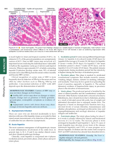
Figure 21.12 Acute viral hepatitis. The predominant histologic changes are: variable degree of necrosis of hepatocytes, most marked in zone
3 (centrilobular); and mononuclear cellular infiltrate in the lobule. Mild degree of liver cell necrosis is seen as ballooning degeneration while
acidophilic Councilman bodies (inbox) are indicative of more severe liver cell injury.
is much higher in Asian and tropical countries (5-20%). An 1. Incubation period: It varies among different hepatotropic
estimated 2-3% of the general population are asymptomatic viruses: for hepatitis A it is about 4 weeks (15-45 days); for
carriers of HCV. Data on HBV carrier state reveal role of 2 hepatitis B the average is 10 weeks (30-180 days); for hepatitis
important factors rendering the individual more vulnerable D about 6 weeks (30-50 days); for hepatitis C the mean
to harbour the organisms—early age at infection and impaired incubation period is about 7 weeks (20-90 days), and for
immunity. Whereas approximately 10% of adults contracting hepatitis E it is 2-8 weeks (15-60 days). The patient remains
hepatitis B infection develop carrier state, 90% of infected asymptomatic during incubation period but the infectivity
neonates fail to clear HBsAg from the serum within 6 months is highest during the last days of incubation period.
SECTION III
and become HBV carriers. 2. Pre-icteric phase: This phase is marked by prodromal
Clinical recognition of carrier state of HBV is more constitutional symptoms that include anorexia, nausea,
frequently done by detection of HBsAg in the serum and less vomiting, fatigue, malaise, distaste for smoking, arthralgia
often by other markers such as HBeAg, HBcAg and and headache. There may be low-grade fever preceding the
antibodies. Concomitant infection of HDV with HBV onset of jaundice, especially in hepatitis A. The earliest
depends upon the demonstration of anti-HD. laboratory evidence of hepatocellular injury in pre-icteric
phase is the elevation of transaminases.
MORPHOLOGIC FEATURES. Carriers of HBV may or 3. Icteric phase: The prodromal period is heralded by the
may not show changes on liver biopsy. onset of clinical jaundice and the constitutional symptoms
Healthy HBV carriers may show no changes or minor diminish. Other features include dark-coloured urine due to
hepatic change such as presence of finely granular, bilirubinuria, clay-coloured stools due to cholestasis, pruritus
Systemic Pathology
ground-glass, eosinophilic cytoplasm as evidence of as a result of elevated serum bile acids, loss of weight and
HBsAg. abdominal discomfort due to enlarged, tender liver. The
Asymptomatic carriers with chronic disease may show diagnosis is based on deranged liver function tests (e.g.
changes of chronic hepatitis and even cirrhosis. elevated levels of serum bilirubin, transaminases and
alkaline phosphatase; prolonged prothrombin time and
II. Asymptomatic Infection hyperglobulinaemia) and serologic detection of hepatitis
These are cases who are detected incidentally to have antigens and antibodies.
infection with one of the hepatitis viruses as revealed by their 4. Post-icteric phase: The icteric phase lasting for about 1
raised serum transaminases or by detection of the presence to 4 weeks is usually followed by clinical and biochemical
of antibodies but are otherwise asymptomatic. recovery in 2 to 12 weeks. The recovery phase is more
prolonged in hepatitis B and hepatitis C. Up to 1% cases of
III. Acute Hepatitis acute hepatitis may develop severe form of the disease
The most common consequence of all hepatotropic viruses (fulminant hepatitis); and 5-10% of cases progress on to
is acute inflammatory involvement of the entire liver. In chronic hepatitis. Evolution into the carrier state (except in
general, type A, B, C, D and E run similar clinical course HAV and HEV infection) has already been described above.
and show identical pathologic findings. MORPHOLOGIC FEATURES. Grossly, the liver is
Clinically, acute hepatitis is categorised into 4 phases:
incubation period, pre-icteric phase, icteric phase and post- slightly enlarged, soft and greenish.
Histologically, the changes are as follows (Fig. 21.12):
icteric phase.