
Page 628 - Textbook of Pathology, 6th Edition
P. 628
612
SECTION III
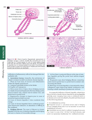
Figure 21.13 Chronic hepatitis. Diagrammatic representation of
pathologic changes in chronic hepatitis (B) contrasted with normal
morphology (A). Photomicrograph on right (C) shows stellate-shaped
portal triad, with extension of fibrous spurs into lobules. The portal tract
is expanded due to increased lymphomononuclear inflammatory cells
which are seen to breach the limiting plate (i.e. hepatocytes at the interface
of portal tract and lobule are destroyed).
infiltration of inflammatory cells in the damaged bile duct i) At first, there is periportal fibrosis at the sites of inter-
epithelial cells. face hepatitis giving the portal tract stellate-shaped
3. Intralobular lesions. Generally, the architecture of appearance.
Systemic Pathology
lobule is retained in mild to moderate chronic hepatitis. ii) Progressive cases show bridging fibrosis connecting
i) There are focal areas of necrosis and inflammation portal tract-to-portal tract or portal tract-to-central vein
within the hepatic parenchyma. traversing the lobule.
ii) Scattered acidophilic bodies in the lobule. iii) End-stage of chronic hepatitis is characterised by dense
iii) Kupffer cell hyperplasia. collagenous septa destroying lobular architecture and
iv) More severe form of injury shows bridging necrosis forming nodules resulting in postnecrotic cirrhosis.
(i.e. bands of necrosed hepatocytes that may bridge portal
tract-to-central vein, central vein-to-central vein, and As prognostic indicator of chronic hepatitis, criteria have
portal tract-to-portal tract). been evolved to classify chronic hepatitis by giving hepatitis
v) Regenerative changes in hepatocytes in cases of activity score (ranging from none to minimal/mild to
persistent hepatocellular necrosis. moderate and severe) described by Knodell and Ishak based
vi) Cases of chronic hepatitis C show moderate fatty on the following features:
change.
vii) Cases of chronic hepatitis B show scattered ground- A. Necroinflammatory activity:
glass hepatocytes indicative of abundance of HBsAg in Periportal necrosis i.e. piecemeal necrosis and/ or bridging
the cytoplasm. necrosis (ranging from score 0 as ‘no necrosis’ to score 4 as
‘multilobular necrosis’).
4. Bridging fibrosis. The onset of fibrosis in chronic Intralobular necrosis, focal or confluent (ranging from score
hepatitis from the area of interface hepatitis and bridging 0 as ‘none’ to score 4 for ‘>10 foci’ for focal necrosis, and
necrosis is a feature of irreversible damage.
score 6 as ‘panacinar/multiacinar’ for confluent necrosis).