
Page 629 - Textbook of Pathology, 6th Edition
P. 629
Extent and depth of portal inflammation (ranging from grade necrosis in which the liver failure is rapid and fulminant 613
0 as ‘no inflammation’ to grade 4 having ‘marked portal occurring in 2-3 weeks.
inflammation’). Fulminant hepatitis of either of the two varieties can occur
from viral and non-viral etiologies:
B. Stage of fibrosis:
Extent and density of fibrosis (ranging from score 0 as ‘no Acute viral hepatitis accounts for about half the cases, most
fibrosis’ to score 6 as ‘cirrhosis’). often from HBV and HCV; less frequently from combined
HBV-HDV and rarely from HAV. However, HEV infection
CLINICAL FEATURES. The clinical features of chronic is a serious complication in pregnant women. In addition,
hepatitis are quite variable ranging from mild disease to full- herpesvirus can also cause serious viral hepatitis.
blown picture of cirrhosis. Non-viral causes include acute hepatitis due to drug
i) Mild chronic hepatitis shows only slight but persistent toxicity (e.g. acetaminophen, non-steroidal anti-
elevation of transaminases (‘transaminitis’) with fatigue, inflammatory drugs, isoniazid, halothane and anti-
malaise and loss of appetite. depressants), poisonings, hypoxic injury and massive
ii) Other cases may show mild hepatomegaly, hepatic infiltration of malignant tumours into the liver.
tenderness and mild splenomegaly. The patients present with features of hepatic failure with
hepatic encephalopathy (page 602). The mortality rate is high
iii) Laboratory findings may reveal prolonged prothrombin if hepatic transplantation is not undertaken.
time, hyperbilirubinaemia, hyperglobulinaemia and
markedly elevated alkaline phosphatase.
MORPHOLOGIC FEATURES. Grossly, the liver is small
iv) Systemic features of circulating immune complexes due and shrunken, often weighing 500-700 gm. The capsule
to HBV and HCV infection may produce features of immune is loose and wrinkled. The sectioned surface shows diffuse
complex vasculitis, glomerulonephritis and cryoglobuli- or random involvement of hepatic lobes. There are
naemia in a proportion of cases. extensive areas of muddy-red and yellow necrosis
However, clinical features do not correlate with morpho- (previously called acute yellow atrophy) and patches of
logic appearance of the liver biopsy. Some patients may have green bile staining.
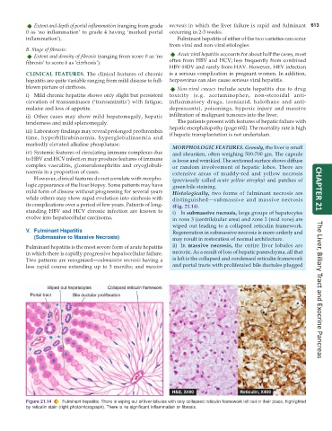
mild form of disease without progressing for several years Histologically, two forms of fulminant necrosis are CHAPTER 21
while others may show rapid evolution into cirrhosis with distinguished—submassive and massive necrosis
its complications over a period of few years. Patients of long- (Fig. 21.14).
standing HBV and HCV chronic infection are known to i) In submassive necrosis, large groups of hepatocytes
evolve into hepatocellular carcinoma. in zone 3 (centrilobular area) and zone 2 (mid zone) are
wiped out leading to a collapsed reticulin framework.
V. Fulminant Hepatitis Regeneration in submassive necrosis is more orderly and
(Submassive to Massive Necrosis) may result in restoration of normal architecture.
Fulminant hepatitis is the most severe form of acute hepatitis ii) In massive necrosis, the entire liver lobules are
in which there is rapidly progressive hepatocellular failure. necrotic. As a result of loss of hepatic parenchyma, all that
Two patterns are recognised—submassive necrosis having a is left is the collapsed and condensed reticulin framework
less rapid course extending up to 3 months; and massive and portal tracts with proliferated bile ductules plugged The Liver, Biliary Tract and Exocrine Pancreas
Figure 21.14 Fulminant hepatitis. There is wiping out of liver lobules with only collapsed reticulin framework left out in their place, highlighted
by reticulin stain (right photomicrograph). There is no significant inflammation or fibrosis.