
Page 631 - Textbook of Pathology, 6th Edition
P. 631
by the spread of Entamoeba histolytica from intestinal lesions. 615
The trophozoite form of amoebae in the colon invade the
colonic mucosa forming flask-shaped ulcers from where they
are carried to the liver in the portal venous system (page
188) . Amoebae multiply and block small intrahepatic portal
radicles resulting in infarction necrosis of the adjacent liver
parenchyma.
The patients, generally from tropical and subtropical
countries, may give history of amoebic dysentery in the past.
Cysts of E. histolytica in stools are present in only 15% of
patients of hepatic amoebiasis. Intermittent low-grade fever,
pain and tenderness in the liver area are common presenting
features. A positive haemagglutination test is quite sensitive
and useful for diagnosis of amoebic liver abscess.
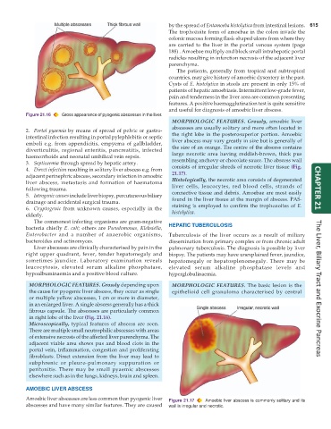
Figure 21.16 Gross appearance of pyogenic abscesses in the liver.
MORPHOLOGIC FEATURES. Grossly, amoebic liver
abscesses are usually solitary and more often located in
2. Portal pyaemia by means of spread of pelvic or gastro-
intestinal infection resulting in portal pylephlebitis or septic the right lobe in the posterosuperior portion. Amoebic
emboli e.g. from appendicitis, empyema of gallbladder, liver abscess may vary greatly in size but is generally of
diverticulitis, regional enteritis, pancreatitis, infected the size of an orange. The centre of the abscess contains
haemorrhoids and neonatal umbilical vein sepsis. large necrotic area having reddish-brown, thick pus
3. Septicaemia through spread by hepatic artery. resembling anchovy or chocolate sauce. The abscess wall
4. Direct infection resulting in solitary liver abscess e.g. from consists of irregular shreds of necrotic liver tissue (Fig.
adjacent perinephric abscess, secondary infection in amoebic 21.17).
liver abscess, metastasis and formation of haematoma Histologically, the necrotic area consists of degenerated
following trauma. liver cells, leucocytes, red blood cells, strands of CHAPTER 21
5. Iatrogenic causes include liver biopsy, percutaneous biliary connective tissue and debris. Amoebae are most easily
drainage and accidental surgical trauma. found in the liver tissue at the margin of abscess. PAS-
6. Cryptogenic from unknown causes, especially in the staining is employed to confirm the trophozoites of E.
elderly. histolytica.
The commonest infecting organisms are gram-negative
bacteria chiefly E. coli; others are Pseudomonas, Klebsiella, HEPATIC TUBERCULOSIS
Enterobacter and a number of anaerobic organisms, Tuberculosis of the liver occurs as a result of miliary
bacteroides and actinomyces. dissemination from primary complex or from chronic adult
Liver abscesses are clinically characterised by pain in the pulmonary tuberculosis. The diagnosis is possible by liver
right upper quadrant, fever, tender hepatomegaly and biopsy. The patients may have unexplained fever, jaundice,
sometimes jaundice. Laboratory examination reveals hepatomegaly or hepatosplenomegaly. There may be
leucocytosis, elevated serum alkaline phosphatase, elevated serum alkaline phosphatase levels and
hypoalbuminaemia and a positive blood culture. hyperglobulinaemia.
MORPHOLOGIC FEATURES. Grossly depending upon MORPHOLOGIC FEATURES. The basic lesion is the
the cause for pyogenic liver abscess, they occur as single epithelioid cell granuloma characterised by central The Liver, Biliary Tract and Exocrine Pancreas
or multiple yellow abscesses, 1 cm or more in diameter,
in an enlarged liver. A single abscess generally has a thick
fibrous capsule. The abscesses are particularly common
in right lobe of the liver (Fig. 21.16).
Microscopically, typical features of abscess are seen.
There are multiple small neutrophilic abscesses with areas
of extensive necrosis of the affected liver parenchyma. The
adjacent viable area shows pus and blood clots in the
portal vein, inflammation, congestion and proliferating
fibroblasts. Direct extension from the liver may lead to
subphrenic or pleuro-pulmonary suppuration or
peritonitis. There may be small pyaemic abscesses
elsewhere such as in the lungs, kidneys, brain and spleen.
AMOEBIC LIVER ABSCESS
Amoebic liver abscesses are less common than pyogenic liver Figure 21.17 Amoebic liver abscess is commonly solitary and its
abscesses and have many similar features. They are caused wall is irregular and necrotic.