
Page 632 - Textbook of Pathology, 6th Edition
P. 632
616 HYDATID DISEASE (ECHINOCOCCOSIS)
Hydatid disease occurs as a result of infection by the larval
cyst stage of the tapeworm, Echinococcus granulosus. The dog
is the common definite host, while man, sheep and cattle
are the intermediate hosts. The dog is infected by eating the
viscera of sheep containing hydatid cysts. The infected faeces
of the dog contaminate grass and farmland from where the
ova are ingested by sheep, pigs and man. Thus, man can
acquire infection by handling dogs as well as by eating conta-
minated vegetables. The ova ingested by man are liberated
from the chitinous wall by gastric juice and pass through
the intestinal mucosa from where they are carried to the liver
by portal venous system. These are trapped in the hepatic
sinusoids where they eventually develop into hydatid cyst.
About 70% of hydatid cysts develop in the liver which acts
as the first filter for ova. However, ova which pass through
the liver enter the right side of the heart and are caught in
the pulmonary capillary bed and form pulmonary hydatid
cysts. Some ova which enter the systemic circulation give
Figure 21.18 Miliary tuberculosis liver. The hepatic parenchyma
shows epithelioid granulomas with small areas of central necrosis and rise to hydatid cysts in the brain, spleen, bone and muscles.
surrounded peripherally by Langhans’ giant cells and lymphocytes. The disease is common in sheep-raising countries such
as Australia, New Zealand and South America. The
uncomplicated hydatid cyst of the liver may be silent or may
caseation necrosis with destruction of the reticulin produce dull ache in the liver area and some abdominal
framework and peripheral cuff of lymphocytes distension.
(Fig. 21.18). Ziehl-Neelsen staining for AFB or culture of Complications of hydatid cyst include its rupture (e.g. into
the organism from the biopsy tissue is confirmatory. Rare the peritoneal cavity, bile ducts and lungs), secondary
lesions consist of tuberculous cholangitis and tuberculous infection and hydatid allergy due to sensitisation of the host
pylephlebitis. with cyst fluid. The diagnosis is made by peripheral blood
SECTION III
eosinophilia, radiologic examination and serologic tests such
as indirect haemagglutination test and Casoni skin test.
Systemic Pathology
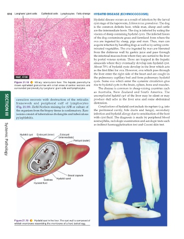
Figure 21.19 Hydatid cyst in the liver. The cyst wall is composed of
whitish membrane resembling the membrane of a hard boiled egg.