
Page 634 - Textbook of Pathology, 6th Edition
P. 634
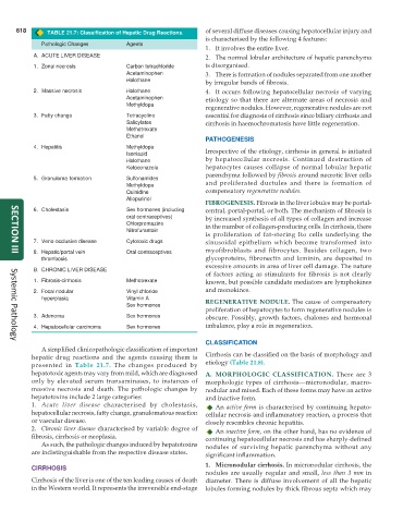
618 TABLE 21.7: Classification of Hepatic Drug Reactions. of several diffuse diseases causing hepatocellular injury and
is characterised by the following 4 features:
Pathologic Changes Agents
1. It involves the entire liver.
A. ACUTE LIVER DISEASE 2. The normal lobular architecture of hepatic parenchyma
1. Zonal necrosis Carbon tetrachloride is disorganised.
Acetaminophen 3. There is formation of nodules separated from one another
Halothane by irregular bands of fibrosis.
2. Massive necrosis Halothane 4. It occurs following hepatocellular necrosis of varying
Acetaminophen etiology so that there are alternate areas of necrosis and
Methyldopa
regenerative nodules. However, regenerative nodules are not
3. Fatty change Tetracycline essential for diagnosis of cirrhosis since biliary cirrhosis and
Salicylates cirrhosis in haemochromatosis have little regeneration.
Methotrexate
Ethanol
PATHOGENESIS
4. Hepatitis Methyldopa Irrespective of the etiology, cirrhosis in general is initiated
Isoniazid
Halothane by hepatocellular necrosis. Continued destruction of
Ketoconazole hepatocytes causes collapse of normal lobular hepatic
parenchyma followed by fibrosis around necrotic liver cells
5. Granuloma formation Sulfonamides
Methyldopa and proliferated ductules and there is formation of
Quinidine compensatory regenerative nodules.
Allopurinol
FIBROGENESIS. Fibrosis in the liver lobules may be portal-
6. Cholestasis Sex hormones (including central, portal-portal, or both. The mechanism of fibrosis is
oral contraceptives) by increased synthesis of all types of collagen and increase
Chlorpromazine in the number of collagen-producing cells. In cirrhosis, there
Nitrofurantoin
is proliferation of fat-storing Ito cells underlying the
7. Veno-occlusive disease Cytotoxic drugs sinusoidal epithelium which become transformed into
8. Hepatic/portal vein Oral contraceptives myofibroblasts and fibrocytes. Besides collagen, two
SECTION III
thrombosis glycoproteins, fibronectin and laminin, are deposited in
excessive amounts in area of liver cell damage. The nature
B. CHRONIC LIVER DISEASE
of factors acting as stimulants for fibrosis is not clearly
1. Fibrosis-cirrhosis Methotrexate known, but possible candidate mediators are lymphokines
2. Focal nodular Vinyl chloride and monokines.
hyperplasia Vitamin A REGENERATIVE NODULE. The cause of compensatory
Sex hormones
proliferation of hepatocytes to form regenerative nodules is
3. Adenoma Sex hormones obscure. Possibly, growth factors, chalones and hormonal
4. Hepatocellular carcinoma Sex hormones imbalance, play a role in regeneration.
CLASSIFICATION
Systemic Pathology
A simplified clinicopathologic classification of important
hepatic drug reactions and the agents causing them is Cirrhosis can be classified on the basis of morphology and
presented in Table 21.7. The changes produced by etiology (Table 21.8).
hepatotoxic agents may vary from mild, which are diagnosed A. MORPHOLOGIC CLASSIFICATION. There are 3
only by elevated serum transaminases, to instances of morphologic types of cirrhosis—micronodular, macro-
massive necrosis and death. The pathologic changes by nodular and mixed. Each of these forms may have an active
hepatotoxins include 2 large categories: and inactive form.
1. Acute liver disease characterised by cholestasis, An active form is characterised by continuing hepato-
hepatocellular necrosis, fatty change, granulomatous reaction cellular necrosis and inflammatory reaction, a process that
or vascular disease. closely resembles chronic hepatitis.
2. Chronic liver disease characterised by variable degree of An inactive form, on the other hand, has no evidence of
fibrosis, cirrhosis or neoplasia. continuing hepatocellular necrosis and has sharply-defined
As such, the pathologic changes induced by hepatotoxins nodules of surviving hepatic parenchyma without any
are indistinguishable from the respective disease states. significant inflammation.
1. Micronodular cirrhosis. In micronodular cirrhosis, the
CIRRHOSIS
nodules are usually regular and small, less than 3 mm in
Cirrhosis of the liver is one of the ten leading causes of death diameter. There is diffuse involvement of all the hepatic
in the Western world. It represents the irreversible end-stage lobules forming nodules by thick fibrous septa which may