
Page 66 - Clinical Anatomy
P. 66
ECA1 7/18/06 6:31 PM Page 51
On the examination of a chest radiograph 51
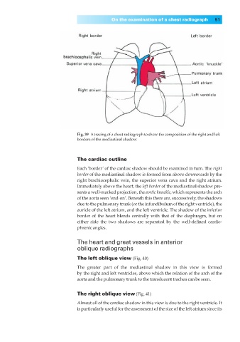
Fig. 39◊Atracing of a chest radiograph to show the composition of the right and left
borders of the mediastinal shadow.
The cardiac outline
Each ‘border’ of the cardiac shadow should be examined in turn. The right
border of the mediastinal shadow is formed from above downwards by the
right brachiocephalic vein, the superior vena cava and the right atrium.
Immediately above the heart, the left border of the mediastinal shadow pre-
sents a well-marked projection, the aortic knuckle, which represents the arch
of the aorta seen ‘end-on’. Beneath this there are, successively, the shadows
due to the pulmonary trunk (or the infundibulum of the right ventricle), the
auricle of the left atrium, and the left ventricle. The shadow of the inferior
border of the heart blends centrally with that of the diaphragm, but on
either side the two shadows are separated by the well-defined cardio-
phrenic angles.
The heart and great vessels in anterior
oblique radiographs
The left oblique view (Fig. 40)
The greater part of the mediastinal shadow in this view is formed
by the right and left ventricles, above which the relation of the arch of the
aorta and the pulmonary trunk to the translucent trachea can be seen.
The right oblique view (Fig. 41)
Almost all of the cardiac shadow in this view is due to the right ventricle. It
is particularly useful for the assessment of the size of the left atrium since its