
Page 29 - untitled
P. 29
AAAC11 21/5/05 10:43 AM Page 28
11 The abdominal wall
Serratus
anterior
Cut edge of external oblique
Linea alba
Linea
semilunaris Cut edge of external oblique
Internal oblique
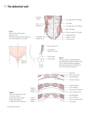
Fig.11.1
Anterior superior iliac spine
Two muscles of the anterior
abdominal wall. Inguinal ligament
The external oblique (on the right) and Superficial Conjoint tendon
the internal oblique (on the left) inguinal ring
Pubic tubercle
A
Rectus abdominis
Deep layer of
superficial fascia
Colles' fascia
Fig.11.2
Fascia penis
The fibrous layer of superficial fascia
Inguinal
can be likened to a pair of bathing trunks
ligament
sewn to the thigh below the inguinal
Dartos ligament and clinging to the penis and
A
muscle scrotum (except for the glans)
Linea alba
A Rectus abdominis
External oblique
Costal cartilages
B
External oblique
Internal oblique
Superior
epigastric Transversus abdominis
artery Transversalis fascia
Fig.11.3
C
Transverse sections through
External oblique
the rectus sheath.
Internal oblique
A: above the costal margin Inferior
B: above the umbilicus epigastric Transversus abdominis
C: above the pubic symphysis artery Peritoneum
28 Abdomen and pelvis