
Page 7 - untitled
P. 7
AAAC01 21/5/05 10:38 AM Page 6
1 The thoracic wall I
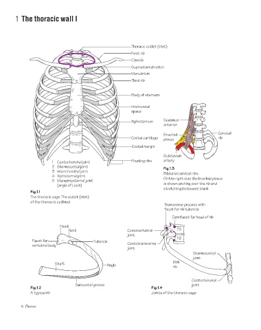
Thoracic outlet (inlet)
First rib
Clavicle
Suprasternal notch
Manubrium
5 Third rib
2
1
Body of sternum
Intercostal
space
4
Xiphisternum Scalenus
anterior
Brachial Cervical
Costal cartilage plexus rib
Costal margin
3
Subclavian
1 Costochondral joint Floating ribs artery
2 Sternocostal joint Fig.1.3
3 Interchondral joint Bilateral cervical ribs.
4 Xiphisternal joint
5 Manubriosternal joint On the right side the brachial plexus
(angle of Louis) is shown arching over the rib and
stretching its lowest trunk
Fig.1.1
The thoracic cage. The outlet (inlet)
of the thorax is outlined
Transverse process with
facet for rib tubercle
Demifacet for head of rib
Head
Neck Costovertebral T5
joint
T6
Facet for Tubercle
vertebral body Costotransverse
joint
Sternocostal
joint
6th
Shaft Angle
rib
Costochondral
Subcostal groove joint
Fig.1.2 Fig.1.4
A typical rib Joints of the thoracic cage
6 Thorax