
Page 1735 - Williams Hematology ( PDFDrive )
P. 1735
1710 Part XI: Malignant Lymphoid Diseases Chapter 105: Plasma Cell Neoplasms: General Considerations 1711
the phosphorylated forms of Paratarg-7 were studied, they were found are not the only driving force in the development of PCN. Although
to have a 6.5-fold higher risk of developing IgM monoclonal gammopa- ethnicity and genealogy affect the prevalence of monoclonal gammo-
thy or macroglobulinemia. The antigen causes continuous autostimula- pathy, they do not impact the rate of progression from monoclonal
76
tion of cognate T-helper cells, which, in turn, specifically activate B cells gammopathy to myeloma. Furthermore, there is a higher incidence in
with high affinity to Paratarg-7. monoclonal gammopathy in relatives of myeloma patients than seen in
the general public. 77,78 These results demonstrate that not only are there
somatic mutations important to the development of PCNs, but genetic
GENETIC ABNORMALITIES IN PLASMA background also contributes to the likelihood of the development of
CELL NEOPLASM plasma cell diseases.
MYELOMA Late Events in Myeloma Progression
Much focus has been placed on risk factors and early genetic events
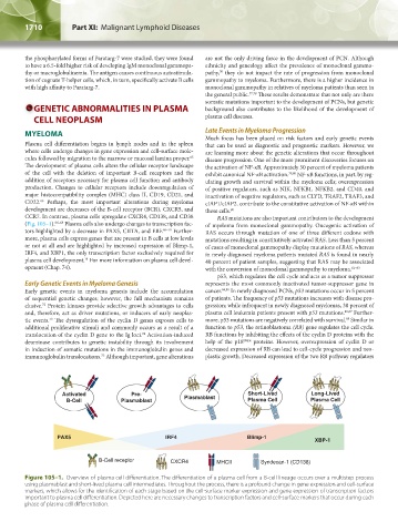
Plasma cell differentiation begins in lymph nodes and in the spleen that can be used as diagnostic and prognostic markers. However, we
where cells undergo changes in gene expression and cell-surface mole- are learning more about the genetic alterations that occur throughout
cules followed by migration to the marrow or mucosal lamina proper. disease progression. One of the more prominent discoveries focuses on
65
The development of plasma cells alters the cellular receptor landscape the activation of NF-κB. Approximately 50 percent of myeloma patients
of the cell with the deletion of important B-cell receptors and the exhibit canonical NF-κB activation. 79,80 NF-κB functions, in part, by reg-
addition of receptors necessary for plasma cell function and antibody ulating growth and survival within the myeloma cells; overexpression
production. Changes to cellular receptors include downregulation of of positive regulators, such as NIK, NFKB1, NFKB2, and CD40, and
major histocompatibility complex (MHC) class II, CD19, CD21, and inactivation of negative regulators, such as CLYD, TRAF2, TRAF3, and
CD22. Perhaps, the most important alterations during myeloma cIAP1/cIAP2, contribute to the constitutive activation of NF-κB within
66
development are decreases of the B-cell receptor (BCR), CXCR5, and these cells. 80
CCR7. In contrast, plasma cells upregulate CXCR4, CD138, and CD38 RAS mutations are also important contributors to the development
(Fig. 105–1). 66–68 Plasma cells also undergo changes to transcription fac- of myeloma from monoclonal gammopathy. Oncogenic activation of
tors highlighted by a decrease in PAX5, CIITA, and EBF. 66–70 Further- RAS occurs through mutation of one of three different codons with
more, plasma cells express genes that are present in B cells at low levels mutations resulting in constitutively activated RAS. Less than 5 percent
or not at all and are highlighted by increased expression of Blimp-1, of cases of monoclonal gammopathy display mutations of RAS, whereas
IRF4, and XBP1, the only transcription factor exclusively required for in newly diagnosed myeloma patients mutated RAS is found in nearly
plasma cell development. For more information on plasma cell devel- 40 percent of patient samples, suggesting that RAS may be associated
71
opment (Chap. 74). with the conversion of monoclonal gammopathy to myeloma. 81–83
p53, which regulates the cell cycle and acts as a tumor suppressor
Early Genetic Events in Myeloma Genesis represents the most commonly inactivated tumor-suppressor gene in
Early genetic events in myeloma genesis include the accumulation cancer. 84,85 In newly diagnosed PCNs, p53 mutations occur in 5 percent
of sequential genetic changes; however, the full mechanism remains of patients. The frequency of p53 mutations increases with disease pro-
elusive. Protein kinases provide selective growth advantages to cells gression; while infrequent in newly diagnosed myelomas, 30 percent of
72
and, therefore, act as driver mutations, or inducers of early neoplas- plasma cell leukemia patients present with p53 mutations. 86,87 Further-
tic events. The dysregulation of the cyclin D genes exposes cells to more, p53 mutations are negatively correlated with survival. Similar in
88
73
additional proliferative stimuli and commonly occurs as a result of a function to p53, the retinoblastoma (RB) gene regulates the cell cycle.
translocation of the cyclin D gene to the Ig loci. Activation-induced RB functions by inhibiting the effects of the cyclin D proteins with the
74
deaminase contributes to genetic instability through its involvement help of the p18 INK4 proteins. However, overexpression of cyclin D or
in induction of somatic mutations in the immunoglobulin genes and decreased expression of RB can lead to cell-cycle progression and neo-
immunoglobulin translocations. Although important, gene alterations plastic growth. Decreased expression of the two RB pathway regulators
75
Activated Pre- Plasmablast Short-Lived Long-Lived
B-Cell Plasmablast Plasma Cell Plasma Cell
PAX5 IRF4 Blimp-1
XBP-1
B-Cell receptor CXCR4 MHCII Syndecan-1 (CD138)
Figure 105–1. Overview of plasma cell differentiation. The differentiation of a plasma cell from a B-cell lineage occurs over a multistep process
using plasmablast and short-lived plasma cell intermediates. Throughout the process, there is a profound change in gene expression and cell-surface
markers, which allows for the identification of each stage based on the cell-surface marker expression and gene expression of transcription factors
important to plasma cell differentiation. Depicted here are necessary changes to transcription factors and cell-surface markers that occur during each
phase of plasma cell differentiation.
Kaushansky_chapter 105_p1707-1720.indd 1710 9/18/15 9:44 AM