
Page 699 - Hall et al (2015) Principles of Critical Care-McGraw-Hill
P. 699
518 PART 4: Pulmonary Disorders
lung tissue tethers open adjacent airways. Ground-glass opacifications dyspnea or radiographic changes. With time, arterial hypoxemia
75
are either absent or minimal. 68 and a widened (a-a)D O 2 are found at rest. In 20% of patients, arterial
Other causes of lower lobe–predominant infiltrates include fibrosis hypoxemia is worse in the upright position and improved with recum-
associated with connective tissue disorders, asbestosis, and chronic bency. This paradoxical pattern is also seen with patent foramen ovale,
73
aspiration. Upper lobe–predominant lesions include sarcoidosis, intrapulmonary arteriovenous malformation, and hepatopulmonary
tuberculosis, fungal infections, silicosis, allergic bronchopulmonary syndrome. Arterial saturation also falls significantly in many patients
aspergillosis, Langerhans cell histiocytosis, ankylosing spondylitis, during REM sleep. 71,73 Sleep-related hypoxemia is due to the exaggerated
berylliosis, cystic fibrosis, and hypersensitivity pneumonitis. If hilar effects of normal nocturnal hypoventilation and V ˙ /Q ˙ variance. Alveolar
adenopathy is present, sarcoidosis, tuberculosis, endemic myco- hypoventilation from respiratory muscle dysfunction or obstructive
ses, malignancy, and berylliosis should be considered. Pleural effu- sleep apnea also may be responsible. 71
sions suggest lymphangioleiomyomatosis, select connective tissue The importance of an anatomic barrier to the diffusion of oxygen (sec-
disorders, asbestos-related lung disease, and drug-induced lung disease. ondary to a thickened, fibrotic interstitium) has been debated. In eight
Extensive parenchymal cysts suggest Langerhans cell histiocytosis, patients with varying types of interstitial lung disease, multiple inert gas
lymphangioleiomyomatosis, and lymphocytic interstitial pneumonia. analysis showed that V ˙ /Q ˙ inequality was the principal defect; diffusion
These conditions can result in diffuse parenchymal infiltrates with limitation contributed to none of the (a-a)D O 2 at rest and only 19% of the
normal or increased lung volumes, as can a mixed process of emphy- (a-a)D O 2 during exercise. However, in 15 patients with IPF also studied
79
sema and IPF. by multiple inert gas elimination, 19% of the (a-a)D O 2 at rest and 40%
■ RESPIRATORY MECHANICS of the (a-a)D O 2 during exercise was attributed to diffusion limitation.
80
V ˙ /Q ˙ inequality remained the principal defect, contributing to 81% of the
–
In end-stage pulmonary fibrosis, pulmonary function tests typically (a-a)D O 2 at rest, and a combination of low Pv from an inadequate cardiac
O 2
show reduced TLC, VC, and IC (see Fig. 58-2). FRC and RV are also output, diffusion limitation, and high V ˙ /Q ˙ variance accounted for the
reduced, though usually to a lesser extent than TLC or VC. Rarely, RV widening of the (a-a)D O 2 during exercise. Intrapulmonary shunt was
is normal when there is early airway closure or decreased elastic recoil small, averaging 2% of cardiac output at rest and 3% during exercise.
pressure at low lung volumes. Both the forced vital capacity (FVC) The dead space to tidal volume ratio (V /V ) may exceed 0.4 (normal
75
t
ds
71
and the forced expiratory volume in 1 second (FEV ) are decreased, but ≤0.3) in end-stage fibrosis. This reflects an increase in the volume of
1
FEV /FVC is generally increased. In this instance, high expiratory flow alveolar dead space and a decrease in tidal volume. When V /V is high,
ds
t
1
rates relative to volume reflect increased elastic recoil pressure. Airway greater minute ventilation is required to maintain alveolar ventilation
resistance is usually normal or low, although reversible and irreversible and a normal Pa CO 2 . Patients may surpass these heightened requirements
obstructive defects do occur. to achieve respiratory alkalosis, perhaps in response to greater afferent
70
Since chest wall compliance and respiratory muscles are normal in stimuli from the fibrotic lung. The development of hypercapnia is an
most patients with pulmonary fibrosis, lung volumes are affected by ominous sign of imminent death.
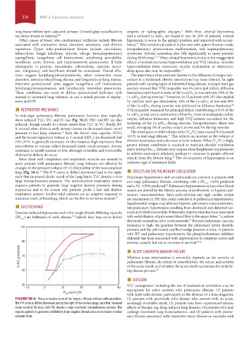
lung (Fig. 58-4). The P-V curve is shifted downward and to the right, ■ EFFECTS ON THE PULMONARY CIRCULATION
changes in the pressure-volume (P-V) relationship of the noncompliant
76
such that increased elastic recoil of the lung limits TLC despite a very Pulmonary hypertension and cor pulmonale are common in patients with
large transpulmonary pressure. The noncompliant respiratory system end-stage pulmonary fibrosis, correlating with a DL <45% predicted
CO
requires patients to generate large negative pleural pressures during and a VC <50% predicted. Pulmonary hypertension occurs when blood
82
inspiration and is the reason why patients prefer a fast and shallow vessels are altered by the fibrotic process, microthrombi, or hypoxic pul-
respiratory pattern. Smaller tidal volumes are an adaptive response to monary vasoconstriction. Since polycythemia and high cardiac output
minimize work of breathing, which can be five to six times normal. 77 are uncommon in IPF, they rarely contribute to pulmonary hypertension.
■ GAS EXCHANGE Supplemental oxygen may alleviate hypoxic pulmonary vasoconstriction,
but pulmonary hypertension resulting from destroyed and distorted vas-
Exercise-induced hypoxemia and a low single-breath diffusing capacity culature is likely irreversible. Pulmonary hypertension has been associated
(DL ) are hallmarks of early disease. Indeed, they may occur before with redistribution of pulmonary blood flow to the upper lobes, a pattern
74
78
CO
that rarely normalizes after corticosteroids. Because pulmonary vascular
81
resistance is high, the gradient between the pulmonary artery diastolic
110
pressure and the pulmonary capillary wedge pressure is wide. In patients
with IPF and pulmonary hypertension, the phosphodiesterase inhibitor
sildenafil has been associated with improvement in symptom scores and
90 exercise capacity but not an increase in survival. 83-85
■
% TLC predicted 70 Whether acute deterioration is reversible depends on the severity of
ACUTE CARDIOPULMONARY FAILURE
pulmonary fibrosis, the extent of comorbidities, the nature and severity
ing disease process.
50 of the acute insult, and whether the acute insult accelerates the underly-
72
■ OUTCOME
30 ICU management including the use of mechanical ventilation may be
–10 0 10 20 30 40 appropriate for select patients with pulmonary fibrosis: (1) patients
P (cm H O) with early/mild disease, particularly in the absence of a firm diagnosis,
2
tm
FIGURE 58-4. Pressure-volume curve of the lung in a 48-year-old man with sarcoidosis. (2) patients with previously mild disease who present with an acute,
The P-V curve is shifted downward and to the right of the normal range, such that increased seemingly reversible insult, (3) patients who have experienced adverse
elastic recoil of the lung limits TLC despite a large maximum transpulmonary pressure. This effects of therapy (eg, drug-induced lung disease), (4) patients who may
requires patients to generate prohibitively large negative pleural pressures to inspire minimal undergo imminent lung transplantation, and (5) patients with pulmo-
amounts of air. nary fibrosis associated with connective tissue disease or vasculitis with
section04.indd 518 1/23/2015 2:20:30 PM