
Page 443 - fbkCardioDiabetes_2017
P. 443
Don’t Let Diabetes Pull Down Our Foot 419
been many treatment options for diabetic ulcer, op-
timal results are yet to be obtained.
There are various strategies in wound dressing to
facilitate wound healing. If the standard treatment
fails to heal the Diabetic foot ulcer, supplementary
and advanced treatment modalities would be re-
quired. They are comparatively effective and have
minimal side effects. They are collagen products
(COL), biological skin equivalents (BSE), biological
dressings (BD), silver products, intermittent pneu-
matic compression therapy (IPC), negative pressure
wound therapy (NPWT), electromagnetic therapy
(EMT), keratinocytes, platelet-derived growth factor
(PDGF), platelet-rich plasma (PRP), hyperbaric oxygen
(HBOT), topical oxygen, Honey dressing and ozone
oxygen etc.
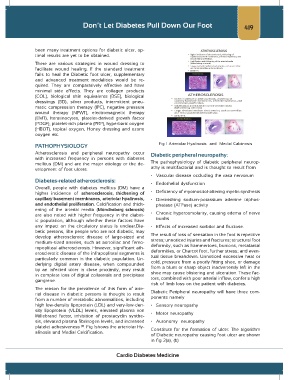
PATHOPHYSIOLOGY Fig 1 Arteriolar Hyalinosis and Medial Calcinosis
Atherosclerosis and peripheral neuropathy occur Diabetic peripheral neuropathy:
with increased frequency in persons with diabetes
mellitus (DM) and are the major etiology or the de- The pathophysiology of diabetic peripheral neurop-
velopment of foot ulcers. athy is multifactorial and is thought to result from
• Vascular disease occluding the vasa nervorum
Diabetes-related atherosclerosis:
• Endothelial dysfunction
Overall, people with diabetes mellitus (DM) have a
higher incidence of atherosclerosis, thickening of • Deficiency of myoinositol-altering myelin synthesis
capillary basement membranes, arteriolar hyalinosis, • Diminishing sodium-potassium adenine triphos-
and endothelial proliferation. Calcification and thick- phatase (ATPase) activity
ening of the arterial media (Mönckeberg sclerosis)
are also noted with higher frequency in the diabet- • Chronic hyperosmolarity, causing edema of nerve
ic population, although whether these factors have trunks
any impact on the circulatory status is unclear.Dia- • Effects of increased sorbitol and fructose.
betic persons, like people who are not diabetic, may
develop atherosclerotic disease of large-sized and The result of loss of sensation in the foot is repetitive
medium-sized arteries, such as aortoiliac and femo- stress; unnoticed injuries and fractures; structural foot
ropopliteal atherosclerosis. However, significant ath- deformity, such as hammertoes, bunions, metatarsal
erosclerotic disease of the infrapopliteal segments is deformities, or Charcot foot, further stress; and even-
particularly common in the diabetic population. Un- tual tissue breakdown. Unnoticed excessive heat or
derlying digital artery disease, when compounded cold, pressure from a poorly fitting shoe, or damage
by an infected ulcer in close proximity, may result from a blunt or sharp object inadvertently left in the
in complete loss of digital collaterals and precipitate shoe may cause blistering and ulceration. These fac-
gangrene. tors, combined with poor arterial inflow, confer a high
risk of limb loss on the patient with diabetes.
The reason for the prevalence of this form of arte-
rial disease in diabetic persons is thought to result Diabetic Peripheral neuropathy will have three com-
from a number of metabolic abnormalities, including ponents namely
high low-density lipoprotein (LDL) and very-low-den- • Sensory neuropathy
sity lipoprotein (VLDL) levels, elevated plasma von • Motor neuropathy
Willebrand factor, inhibition of prostacyclin synthe-
sis, elevated plasma fibrinogen levels, and increased • Autonomy neuropathy
platelet adhesiveness . Fig 1shows the arteriolar Hy- Constitute for the formation of ulcer. The algorithm
(2)
alinosis and Medial Calcification.
of Diabetic neuropathy causing foot ulcer are shown
in fig 2(a), (b)
Cardio Diabetes Medicine