
Page 333 - Color_Atlas_of_Physiology_5th_Ed._-_A._Despopoulos_2003
P. 333
Unlike monosynaptic stretch reflexes, poly-
Polysynaptic Reflexes
synaptic reflexes occur through the co-activa-
Unlike proprioceptive reflexes (! p. 316), tion of α and γ motoneurons (! p. 316). The re-
polysynaptic reflexes are activated by sensors flex excitability of α motoneurons is largely
that are spatially separate from the effector controlled by supraspinal centers via multiple
organ. This type of reflex is called polysynaptic, interneurons (! p. 324). The brain can there-
since the reflex arc involves many synapses in fore shorten the reflex time of spinal cord re-
flexes when a noxious stimulus is anticipated.
Central Nervous System and Senses in nose _! sneezing. The response spreads of reflexes (hyperreflexia) and stereotypic reflexes.
series. This results in a relatively long reflex
time. The intensity of the response is depend-
Supraspinal lesions or interruption of descending
ent on the duration and intensity of stimulus,
tracts (e.g., in paraplegics) can lead to exaggeration
which is temporally and spatially summated in
the CNS (! p. 52). Example: itching sensation
The absence of reflexes (areflexia) corresponds to
specific disorders of the spinal cord or peripheral
nerve.
when the stimulus intensity increases (e.g.,
coughing ! choking cough). Protective reflexes
(e.g., withdrawal reflex, corneal and lacrimal
Synaptic Inhibition
reflexes, coughing and sneezing), nutrition re-
GABA (γ-aminobutyric acid) and glycine
flexes (e.g., swallowing, sucking reflexes), loco-
in the spinal cord. Presynaptic inhibition (! B)
flexes are polysynaptic reflexes. Certain re-
occurs frequently in the CNS, for example, at
flexes, e.g., plantar reflex, cremasteric reflex
synapses between type Ia afferents and α mo-
and abdominal reflex, are used as diagnostic
12 motor reflexes, and the various autonomic re- (! p. 55f.) function as inhibitory transmitters
toneurons, and involves axoaxonic synapses of
tests.
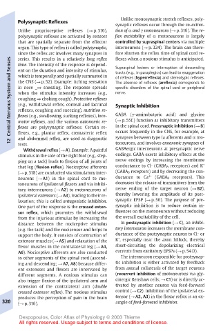
Withdrawal reflex (! A). Example: A painful GABAergic interneurons at presynaptic nerve
stimulus in the sole of the right foot (e.g., step- endings. GABA exerts inhibitory effects at the
ping on a tack) leads to flexion of all joints of nerve endings by increasing the membrane
–
that leg (flexion reflex). Nociceptive afferents conductance to Cl (GABA A receptors) and K +
(! p. 318) are conducted via stimulatory inter- (GABA B receptors) and by decreasing the con-
2+
neurons (! A1) in the spinal cord to mo- ductance to Ca (GABA B receptors). This
toneurons of ipsilateral flexors and via inhibi- decreases the release of transmitters from the
tory interneurons (! A2) to motoneurons of nerve ending of the target neuron (! B2),
ipsilateral extensors (! A3), leading to their re- thereby lowering the amplitude of its post-
laxation; this is called antagonistic inhibition. synaptic EPSP (! p. 50). The purpose of pre-
One part of the response is the crossed exten- synaptic inhibition is to reduce certain in-
sor reflex, which promotes the withdrawal fluences on the motoneuron without reducing
from the injurious stimulus by increasing the the overall excitability of the cell.
distance between the nociceptive stimulus In postsynaptic inhibition (! C), an inhibi-
(e.g. the tack) and the nocisensor and helps to tory interneuron increases the membrane con-
–
support the body. It consists of contraction of ductance of the postsynaptic neuron to Cl or
+
extensor muscles (! A5) and relaxation of the K , especially near the axon hillock, thereby
flexor muscles in the contralateral leg (! A4, short-circuiting the depolarizing electrical
A6). Nociceptive afferents are also conducted currents from excitatory EPSPs (! p. 54 D).
to other segments of the spinal cord (ascend- The interneuron responsible for postsynap-
ing and descending; ! A7, A8) because differ- tic inhibition is either activated by feedback
ent extensors and flexors are innervated by from axonal collaterals of the target neurons
different segments. A noxious stimulus can (recurrent inhibition of motoneurons via gly-
also trigger flexion of the ipsilateral arm and cinergic Renshaw cells; ! C1) or is directly ac-
extension of the contralateral arm (double tivated by another neuron via feed-forward
crossed extensor reflex). The noxious stimulus control (! C2). Inhibition of the ipsilateral ex-
produces the perception of pain in the brain tensor (! A2, A3) in the flexor reflex is an ex-
320 (! p. 316). ample of feed-forward inhibition.
Despopoulos, Color Atlas of Physiology © 2003 Thieme
All rights reserved. Usage subject to terms and conditions of license.