
Page 343 - Clinical Anatomy
P. 343
ECA5 7/18/06 6:51 PM Page 328
328 The head and neck
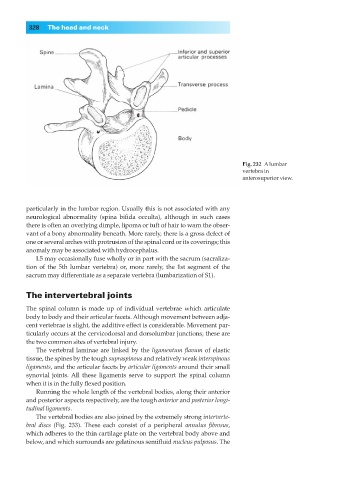
Fig. 232◊Alumbar
vertebra in
anterosuperior view.
particularly in the lumbar region. Usually this is not associated with any
neurological abnormality (spina bifida occulta), although in such cases
there is often an overlying dimple, lipoma or tuft of hair to warn the obser-
vant of a bony abnormality beneath. More rarely, there is a gross defect of
one or several arches with protrusion of the spinal cord or its coverings; this
anomaly may be associated with hydrocephalus.
L5 may occasionally fuse wholly or in part with the sacrum (sacraliza-
tion of the 5th lumbar vertebra) or, more rarely, the 1st segment of the
sacrum may differentiate as a separate vertebra (lumbarization of S1).
The intervertebral joints
The spinal column is made up of individual vertebrae which articulate
body to body and their articular facets. Although movement between adja-
cent vertebrae is slight, the additive effect is considerable. Movement par-
ticularly occurs at the cervicodorsal and dorsolumbar junctions; these are
the two common sites of vertebral injury.
The vertebral laminae are linked by the ligamentum flavum of elastic
tissue, the spines by the tough supraspinous and relatively weak interspinous
ligaments, and the articular facets by articular ligaments around their small
synovial joints. All these ligaments serve to support the spinal column
when it is in the fully flexed position.
Running the whole length of the vertebral bodies, along their anterior
and posterior aspects respectively, are the tough anterior and posterior longi-
tudinal ligaments.
The vertebral bodies are also joined by the extremely strong interverte-
bral discs (Fig. 233). These each consist of a peripheral annulus fibrosus,
which adheres to the thin cartilage plate on the vertebral body above and
below, and which surrounds are gelatinous semifluid nucleus pulposus. The