Page 344 - Clinical Anatomy
P. 344

ECA5  7/18/06  6:51 PM  Page 329


                                                                       The vertebral column    329
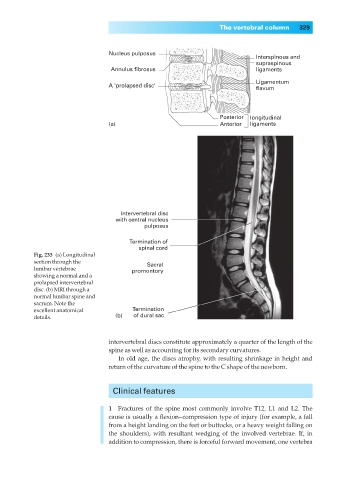


                                        Nucleus pulposus
                                                                                  Interspinous and
                                                                                  supraspinous
                                        Annulus fibrosus                          ligaments
                                                                                  Ligamentum
                                        A 'prolapsed disc'
                                                                                  flavum


                                                                        Posterior  longitudinal
                                        (a)                             Anterior  ligaments


                                           Intervertebral disc
                                         with central nucleus
                                                  pulposus

                                             Termination of
                                                spinal cord
                  Fig. 233◊(a) Longitudinal
                  section through the              Sacral
                  lumbar vertebrae            promontory
                  showing a normal and a
                  prolapsed intervertebral
                  disc. (b) MRI through a
                  normal lumbar spine and
                  sacrum. Note the
                  excellent anatomical        Termination
                  details.               (b)   of dural sac


                                       intervertebral discs constitute approximately a quarter of the length of the
                                       spine as well as accounting for its secondary curvatures.
                                          In old age, the discs atrophy, with resulting shrinkage in height and
                                       return of the curvature of the spine to the C shape of the newborn.


                                         Clinical features


                                       1◊◊Fractures of the spine most commonly involve T12, L1 and L2. The
                                       cause is usually a flexion–compression type of injury (for example, a fall
                                       from a height landing on the feet or buttocks, or a heavy weight falling on
                                       the shoulders), with resultant wedging of the involved vertebrae. If, in
                                       addition to compression, there is forceful forward movement, one vertebra
   339   340   341   342   343   344   345   346   347   348   349