
Page 369 - Clinical Anatomy
P. 369
ECA6 7/18/06 6:54 PM Page 354
354 The central nervous system
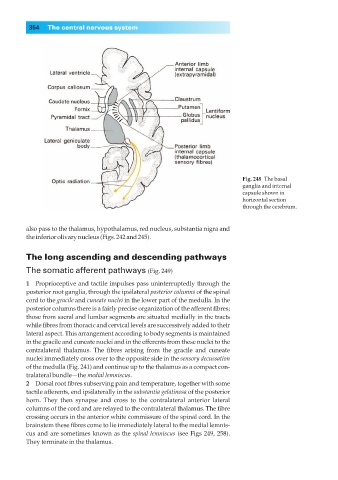
Fig. 248◊The basal
ganglia and internal
capsule shown in
horizontal section
through the cerebrum.
also pass to the thalamus, hypothalamus, red nucleus, substantia nigra and
the inferior olivary nucleus (Figs. 242 and 245).
The long ascending and descending pathways
The somatic afferent pathways (Fig. 249)
1◊◊Proprioceptive and tactile impulses pass uninterruptedly through the
posterior root ganglia, through the ipsilateral posterior columns of the spinal
cord to the gracile and cuneate nuclei in the lower part of the medulla. In the
posterior columns there is a fairly precise organization of the afferent fibres;
those from sacral and lumbar segments are situated medially in the tracts
while fibres from thoracic and cervical levels are successively added to their
lateral aspect. This arrangement according to body segments is maintained
in the gracile and cuneate nuclei and in the efferents from these nuclei to the
contralateral thalamus. The fibres arising from the gracile and cuneate
nuclei immediately cross over to the opposite side in the sensory decussation
of the medulla (Fig. 241) and continue up to the thalamus as a compact con-
tralateral bundle—the medial lemniscus.
2◊◊Dorsal root fibres subserving pain and temperature, together with some
tactile afferents, end ipsilaterally in the substantia gelatinosa of the posterior
horn. They then synapse and cross to the contralateral anterior lateral
columns of the cord and are relayed to the contralateral thalamus. The fibre
crossing occurs in the anterior white commissure of the spinal cord. In the
brainstem these fibres come to lie immediately lateral to the medial lemnis-
cus and are sometimes known as the spinal lemniscus (see Figs 249, 258).
They terminate in the thalamus.