
Page 7 - The Netter Collection of Medical Illustrations - Integumentary System_ Volume 4 ( PDFDrive )
P. 7
ABOUT THE SERIES
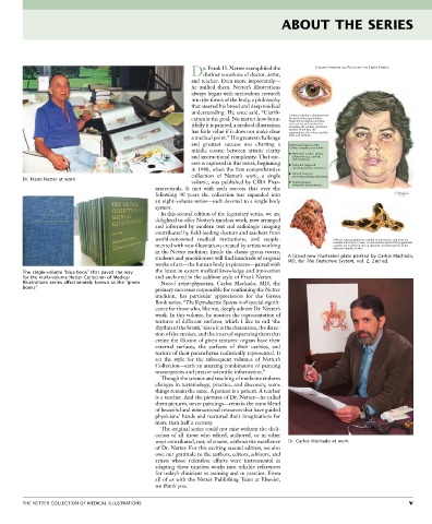
r. Frank H. Netter exemplified the CUSHING’S SYNDROME IN A PATIENT WITH THE CARNEY COMPLEX
Ddistinct vocations of doctor, artist,
and teacher. Even more importantly—
he unified them. Netter’s illustrations
always began with meticulous research
into the forms of the body, a philosophy
that steered his broad and deep medical
understanding. He once said, “Clarifi
cation is the goal. No matter how beau Carney complex is characterized
by spotty skin pigmentation.
Pigmented lentigines and blue
tifully it is painted, a medical illustration nevi can be seen on the face–
including the eyelids, vermillion
has little value if it does not make clear borders of the lips, the
conjunctivae, the sclera–and the
a medical point.” His greatest challenge labia and scrotum.
and greatest success was charting a Additional features of the
middle course between artistic clarity Carney complex can include:
and instructional complexity. That suc Myxomas: cardiac atrium,
cutaneous (e.g., eyelid),
and mammary
cess is captured in this series, beginning Testicular large-cell
in 1948, when the first comprehensive calcifying Sertoli cell tumors
collection of Netter’s work, a single Growth-hormone
secereting pituitary adenomas
Dr. Frank Netter at work volume, was published by CIBA Phar Psammomatous
maceuticals. It met with such success that over the melanotic schwannomas
following 40 years the collection was expanded into
an eightvolume series—each devoted to a single body
system.
In this second edition of the legendary series, we are
delighted to offer Netter’s timeless work, now arranged
and informed by modern text and radiologic imaging
contributed by fieldleading doctors and teachers from
worldrenowned medical institutions, and supple PPNAD adrenal glands are usually of normal size and most are
mented with new illustrations created by artists working studded with black, brown, or red nodules. Most of the pigmented
nodules are less than 4 mm in diameter and interspersed in the
in the Netter tradition. Inside the classic green covers, adjacent atrophic cortex.
students and practitioners will find hundreds of original A brand new illustrated plate painted by Carlos Machado,
works of art—the human body in pictures—paired with MD, for The Endocrine System, vol. 2, 2nd ed.
The single-volume “blue book” that paved the way the latest in expert medical knowledge and innovation
for the multi-volume Netter Collection of Medical and anchored in the sublime style of Frank Netter.
Illustrations series affectionately known as the “green Noted artistphysician, Carlos Machado, MD, the
books” primary successor responsible for continuing the Netter
tradition, has particular appreciation for the Green
Book series. “The Reproductive System is of special signifi
cance for those who, like me, deeply admire Dr. Netter’s
work. In this volume, he masters the representation of
textures of different surfaces, which I like to call ‘the
rhythm of the brush,’ since it is the dimension, the direc
tion of the strokes, and the interval separating them that
create the illusion of given textures: organs have their
external surfaces, the surfaces of their cavities, and
texture of their parenchyma realistically represented. It
set the style for the subsequent volumes of Netter’s
Collection—each an amazing combination of painting
masterpieces and precise scientific information.”
Though the science and teaching of medicine endures
changes in terminology, practice, and discovery, some
things remain the same. A patient is a patient. A teacher
is a teacher. And the pictures of Dr. Netter—he called
them pictures, never paintings—remain the same blend
of beautiful and instructional resources that have guided
physicians’ hands and nurtured their imaginations for
more than half a century.
The original series could not exist without the dedi
cation of all those who edited, authored, or in other
ways contributed, nor, of course, without the excellence Dr. Carlos Machado at work
of Dr. Netter. For this exciting second edition, we also
owe our gratitude to the authors, editors, advisors, and
artists whose relentless efforts were instrumental in
adapting these timeless works into reliable references
for today’s clinicians in training and in practice. From
all of us with the Netter Publishing Team at Elsevier,
we thank you.
THE NETTER COLLECTION OF MEDICAL ILLUSTRATIONS v