
Page 141 - AACN Essentials of Critical-Care Nursing Pocket Handbook, Second Edition
P. 141
128
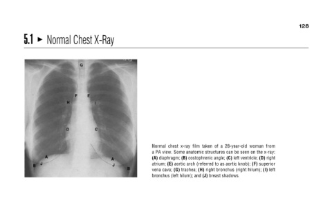
Normal chest x-ray film taken of a 28-year-old woman from
a PA view. Some anatomic structures can be seen on the x-ray:
superior left
aortic arch (referred to as aortic knob); (F) (A) diaphragm; (B) costophrenic angle; (C) left ventricle; (D) right right bronchus (right hilum); (I) (H) bronchus (left hilum); and (J) breast shadows.
(E) trachea; vena cava; (G)
atrium;
Normal Chest X-Ray
5.1