
Page 143 - untitled
P. 143
AAAC64 21/5/05 11:02 AM Page 142
64 The oesophagus and trachea and the thyroid gland
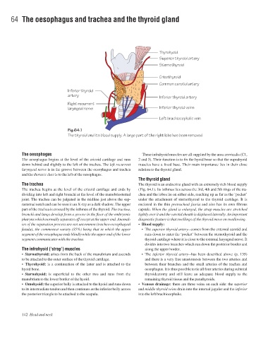
Thyrohyoid
Superior thyroid artery
Sternothyroid
Cricothyroid
Common carotid artery
Inferior thyroid
artery
Inferior thyroid artery
Right recurrent
laryngeal nerve Inferior thyroid veins
Left brachiocephalic vein
Fig.64.1
The thyroid and its blood supply. A large part of the right lobe has been removed
The oesophagus These infrahyoid muscles are all supplied by the ansa cervicalis (C1,
The oesophagus begins at the level of the cricoid cartilage and runs 2 and 3). Their function is to fix the hyoid bone so that the suprahyoid
down behind and slightly to the left of the trachea. The left recurrent muscles have a fixed base. Their main importance lies in their close
laryngeal nerve is in the groove between the oesophagus and trachea relation to the thyroid gland.
and the thoracic duct is to the left of the oesophagus.
The thyroid gland
The trachea The thyroid is an endocrine gland with an extremely rich blood supply
The trachea begins at the level of the cricoid cartilage and ends by (Fig. 64.1). Its isthmus lies across the 3rd, 4th and 5th rings of the tra-
dividing into left and right bronchi at the level of the manubriosternal chea and the lobes lie on either side, reaching up as far as the ‘pocket’
joint. The trachea can be palpated in the midline just above the sup- under the attachment of sternothyroid to the thyroid cartilage. It is
rasternal notch and can be seen in an X-ray as a dark shadow. The upper enclosed in the thin pretracheal fascia and also has its own fibrous
part of the trachea is crossed by the isthmus of the thyroid. The trachea, capsule. When the gland is enlarged, the strap muscles are stretched
bronchi and lungs develop from a groove in the floor of the embryonic tightly over it and the carotid sheath is displaced laterally. An important
pharynx which normally separates off except at the upper end. Anomal- diagnostic feature is that swellings of the thyroid move on swallowing.
ies of the separation process are not uncommon (tracheo-oesophageal • Blood supply:
fistula), the commonest variety (85%) being that in which the upper •The superior thyroid arteryacomes from the external carotid and
segment of the oesophagus ends blindly while the upper end of the lower runs down to enter the ‘pocket’ between the sternothyroid and the
segment communicates with the trachea. thyroid cartilage where it is close to the external laryngeal nerve. It
divides into two branches which run down the posterior border and
The infrahyoid (‘strap’) muscles along the upper border.
• Sternothyroid: arises from the back of the manubrium and ascends •The inferior thyroid arteryahas been described above (p. 135)
to be attached to the outer surface of the thyroid cartilage. and there is a very free anastomosis between the two arteries and
• Thyrohyoid: is a continuation of the latter and is attached to the between their branches and the small arteries of the trachea and
hyoid bone. oesophagus. It is thus possible to tie all four arteries during subtotal
• Sternohyoid: is superficial to the other two and runs from the thyroidectomy and still leave an adequate blood supply to the
manubrium to the lower border of the hyoid. remaining thyroid tissue and the parathyroids.
• Omohyoid: the superior belly is attached to the hyoid and runs down • Venous drainage: there are three veins on each side: the superior
to its intermediate tendon and then continues as the inferior belly across and middle thyroid veins drain into the internal jugular and the inferior
the posterior triangle to be attached to the scapula. into the left brachiocephalic.
142 Head and neck