Page 23 - untitled
P. 23

AAAC08  23/05/2005  3:06 PM  Page 22
            8 The heart II


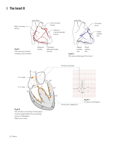
                                                           Left coronary                                   Coronary
                                                           artery                                          sinus
                     Right coronary
                     artery
                                                               Anterior
                                                               interventricular                              Great
                                                               branch                                        cardiac
                                                                                                             vein


                                              Marginal  Posterior                       Middle  Small
                     Fig.8.1                  artery    interventricular                cardiac  cardiac
                     The coronary arteries.             branch                          vein    vein
                     Variations are common                                     Fig.8.2
                                                                               The venous drainage of the heart


                                                                       Atrial conduction


                         S–A node
                                                          65


                         A–V node                 0
                                                               50
                                  55                                                      P         T
                                                                                            QRS
                                                         15      40

                                                                                                Fig.8.4
                                                12
                                         35                                                     An electrocardiogram
                                                                       Ventricular conduction
                                                  25         35
                     Fig.8.3
                     The direction and timing of the spread
                     of action potential in the conducting
                     system of the heart.
                     Times are in msec


                22 Thorax
   18   19   20   21   22   23   24   25   26   27   28