
Page 37 - untitled
P. 37
AAAC14 21/5/05 10:42 AM Page 36
14 The peritoneum
Subphrenic space
Diaphragm
Epiploic foramen (of Winslow)
Upper recess of
Portal vein
omental bursa
Inferior vena cava
Liver
Aorta
Lesser omentum
Epiploic foramen Left kidney
(in the distance)
Splenic artery
Omental bursa
Pancreas Lienorenal ligament
Stomach Spleen
Transverse mesocolon Short gastric
Duodenum (third part) vessels
Transverse colon Gastrosplenic
Small intestine ligament
Stomach
Mesentery
Lesser omentum
Greater omentum Hepatic artery
Fusion between layers Common bile duct
of greater omentum Liver
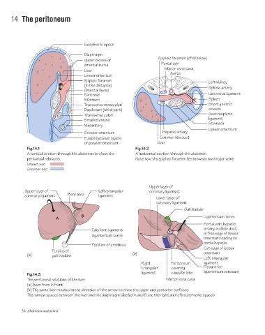
Fig.14.1 Fig.14.2
A vertical section through the abdomen to show the A horizontal section through the abdomen.
peritoneal relations. Note how the epiploic foramen lies between two major veins
Lesser sac
Greater sac
Upper layer of
Upper layer of Left triangular coronary ligament
coronary ligament Bare area ligament
Lower layer of
coronary ligament
Gall bladder
B Ligamentum teres
A
Portal vein, hepatic
Falciform ligament artery and bile duct
in free edge of lesser
Ligamentum teres
omentum leading to
porta hepatis
Position of umbilicus
Cut edge of lesser
Fundus of omentum
(a) gall bladder (b)
Left triangular
Right Peritoneum ligament
triangular covering Fissure for
ligament caudate lobe ligamentum venosum
Fig.14.3
The peritoneal relations of the liver Inferior vena cava
(a) Seen from in front
(b) The same liver rotated in the direction of the arrow to show the upper and posterior surfaces.
The narrow spaces between the liver and the diaphragm labelled A and B are the right and left subphrenic spaces
36 Abdomen and pelvis