
Page 267 - ACCCN's Critical Care Nursing
P. 267
244 P R I N C I P L E S A N D P R A C T I C E O F C R I T I C A L C A R E
● vascular signs: arterial emboli, intracranial haemor- Artery
rhage, Janeway lesions (erythematous spots on the
palms and feet) or conjunctival haemorrhages
● immunological signs: Osler nodes (painful, red-
dened nodules on the fingers and the feet), or
glomerulonephritis 98
A
Echocardiography may reveal vegetations, abscess and Fusiform area
valvular abnormalities, but endocarditis is more a clinical Artery
diagnosis based on the appearance of febrile illness, posi-
tive blood cultures with organisms known to cause endo-
carditis, new murmur and vascular features.
Management
B Sacculated area
Prosthetic valve endocarditis must be aggressively
100
managed, as mortality may be as high as 65%. Impaired
valvular opening, even obstruction, may occur or the Torn intima
100
prosthetic valve may become unseated. Reoperation to False
replace the affected valve should be undertaken when Blood channel
valvular dysfunction is present. Antibiotic therapy is pro- flow created
vided empirically until blood culture and sensitivities are
established. Cardiac failure, if present, is managed along C
standard lines (see section on Nursing management of Ruptured area with
acute heart failure). Observations during endocarditis clot covering the
should be directed at detecting embolic complications opening
involving the brain, kidneys, or spleen; development and
progress of heart failure; progress of the febrile illness, Blood
flow
including hydration and dietary status.
D
Prophylactic antibiotic coverage should be undertaken
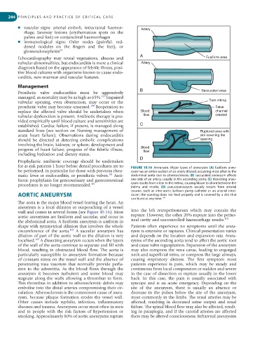
for at-risk patients 1 hour before dental procedures are to FIGURE 10.16 Aneurysm. Major types of aneurysm: (A) fusiform aneu-
be performed, in particular for those with previous rheu- rysm has an entire section of an artery dilated, occurring most often in the
101
matic fever or endocarditis, or prosthetic valves. Anti- abdominal aorta due to atherosclerosis; (B) sacculated aneurysm affects
biotic prophylaxis for genitourinary and gastrointestinal one side of an artery, usually in the ascending aorta; (C) dissecting aneu-
procedures is no longer recommended. 101 rysm results from a tear in the intima, causing blood to shunt between the
intima and media; (D) pseudoaneurysm usually results from arterial
trauma, such as intra-aortic balloon pump catheter or an arterial intro-
AORTIC ANEURYSM ducer; the opening does not heal properly and is covered by a clot that
106
can burst at any time.
The aorta is the major blood vessel leaving the heart. An
aneurysm is a local dilation or outpouching of a vessel
wall and comes in several forms (see Figure 10.16). Most into the left retroperitoneum which may contain the
aortic aneurysms are fusiform and saccular, and occur in rupture. However, the other 20% rupture into the perito-
102
the abdominal aorta. A fusiform aneurysm is uniform in neal cavity and uncontrolled haemorrhage results.
shape with symmetrical dilation that involves the whole Patients often experience no symptoms until the aneu-
102
circumference of the aorta. A saccular aneurysm has rysm is extensive or ruptures. Clinical presentation varies
dilation of part of the aortic wall so the dilation is very and depends on the location and expansion rate. Aneu-
102
localised. A dissecting aneurysm occurs when the layers rysms of the ascending aorta tend to affect the aortic root
of the wall of the aorta continue to separate and fill with and cause valve regurgitation. Expansion of the aneurysm
blood, resulting in obstructed blood flow. The aorta is may also compress the vena cavae, leading to engorged
particularly susceptible to aneurysm formation because neck and superficial veins, or compress the large airways,
of constant stress on the vessel wall and the absence of causing respiratory distress. The first symptom most
penetrating vasa vasorum that normally provide perfu- patients experience is pain, which may be steady and
sion to the adventitia. As the blood flows through the continuous from local compression or sudden and severe
aneurysm it becomes turbulent and some blood may in the case of dissection or rupture usually in the lower
stagnate along the walls allowing a thrombus to form. back. In this case, the pain is usually associated with
This thrombus in addition to atherosclerotic debris may syncope and is an acute emergency. Depending on the
embolise into the distal arteries compromising their cir- site of the aneurysm, there is usually an absence or
culation. Atherosclerosis is the commonest cause of aneu- decrease in the pulses below the site of the aneurysm,
rysm, because plaque formation erodes the vessel wall. most commonly in the limbs. The renal arteries may be
Other causes include syphilis, infection, inflammatory affected, resulting in decreased urine output and renal
diseases and trauma. Aneurysms occur most often in men failure. The spinal blood flow may also be affected, result-
and in people with the risk factors of hypertension or ing in paraplegia, and if the carotid arteries are affected
smoking. Approximately 80% of aortic aneurysms rupture there may be altered consciousness. Infrarenal aneurysms