
Page 758 - ACCCN's Critical Care Nursing
P. 758
Pregnancy and Postpartum Considerations 735
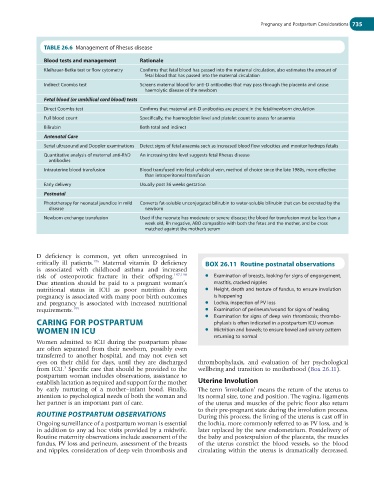
TABLE 26.6 Management of Rhesus disease
Blood tests and management Rationale
Kleihauer-Betke test or flow cytometry Confirms that fetal blood has passed into the maternal circulation, also estimates the amount of
fetal blood that has passed into the maternal circulation
Indirect Coombs test Screens maternal blood for anti-D antibodies that may pass through the placenta and cause
haemolytic disease of the newborn
Fetal blood (or umbilical cord blood) tests
Direct Coombs test Confirms that maternal anti-D antibodies are present in the fetal/newborn circulation
Full blood count Specifically, the haemoglobin level and platelet count to assess for anaemia
Bilirubin Both total and indirect
Antenatal Care
Serial ultrasound and Doppler examinations Detect signs of fetal anaemia such as increased blood flow velocities and monitor hydrops fetalis
Quantitative analysis of maternal anti-RhD An increasing titre level suggests fetal Rhesus disease
antibodies
Intrauterine blood transfusion Blood transfused into fetal umbilical vein, method of choice since the late 1980s, more effective
than intraperitoneal transfusion
Early delivery Usually post 36 weeks gestation
Postnatal
Phototherapy for neonatal jaundice in mild Converts fat-soluble unconjugated bilirubin to water-soluble bilirubin that can be excreted by the
disease newborn
Newborn exchange transfusion Used if the neonate has moderate or severe disease; the blood for transfusion must be less than a
week old, Rh negative, ABO compatible with both the fetus and the mother, and be cross
matched against the mother’s serum
D deficiency is common, yet often unrecognised in
critically ill patients. 196 Maternal vitamin D deficiency BOX 26.11 Routine postnatal observations
is associated with childhood asthma and increased
risk of osteoporotic fracture in their offspring. 197,198 l Examination of breasts, looking for signs of engorgement,
Due attention should be paid to a pregnant woman’s mastitis, cracked nipples
nutritional status in ICU as poor nutrition during l Height, depth and texture of fundus, to ensure involution
pregnancy is associated with many poor birth outcomes is happening
and pregnancy is associated with increased nutritional l Lochia, inspection of PV loss
requirements. 199 l Examination of perineum/wound for signs of healing
l Examination for signs of deep vein thrombosis; thrombo-
CARING FOR POSTPARTUM phylaxis is often indicated in a postpartum ICU woman
WOMEN IN ICU l Mictrition and bowels; to ensure bowel and urinary pattern
returning to normal
Women admitted to ICU during the postpartum phase
are often separated from their newborn, possibly even
transferred to another hospital, and may not even set
eyes on their child for days, until they are discharged thrombophylaxis, and evaluation of her psychological
3
from ICU. Specific care that should be provided to the wellbeing and transition to motherhood (Box 26.11).
postpartum woman includes observations, assistance to
establish lactation as required and support for the mother Uterine Involution
by early nurturing of a mother–infant bond. Finally, The term ‘involution’ means the return of the uterus to
attention to psychological needs of both the woman and its normal size, tone and position. The vagina, ligaments
her partner is an important part of care. of the uterus and muscles of the pelvic floor also return
to their pre-pregnant state during the involution process.
ROUTINE POSTPARTUM OBSERVATIONS During this process, the lining of the uterus is cast off in
Ongoing surveillance of a postpartum woman is essential the lochia, more commonly referred to as PV loss, and is
in addition to any ad hoc visits provided by a midwife. later replaced by the new endometrium. Postdelivery of
Routine maternity observations include assessment of the the baby and postexpulsion of the placenta, the muscles
fundus, PV loss and perineum, assessment of the breasts of the uterus constrict the blood vessels, so the blood
and nipples, consideration of deep vein thrombosis and circulating within the uterus is dramatically decreased.