


Page 80 - Concise Pathology for Exam Preparation ( PDFDrive )
P. 80
3  Healing and Repair  65


                            Formation of a haematoma due to bleeding from torn blood vessels


                                Loose meshwork formed by blood fills the fracture gap

                                        Local inflammatory response


                          Exudation of fibrin, polymorphs and macrophages at the site of fracture


                             Ingrowth of granulation tissue (begins with neovascularization)


               The degranulated platelets and inflammatory cells secrete mediators that activate osteoprogenitor cells in the
                               periosteum, medullary cavity and surrounding soft tissue

                            Soft tissue callus or procallus formation (end of the first week)


                      Osteoprogenitor cells lay down collagen as well as osteoid matrix in granulation tissue


                             •  Calcification of osteoid (woven bone callus)
                             •  Deposition of fibrocartilage and hyaline cartilage


               Woven bone is cleared away by osteoclasts and the calcified cartilage disintegrates; in its place lamellar bone
                                      with haversian system is laid down


                                             Remodelling
                       (realignment and mechanical chopping of bone; development of medullary canal)


                                         Compact bone formation
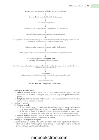
                                FLOWCHART 3.6.  Steps in callus formation.


               2.  Healing of nervous tissue:
                 (a)  Central nervous system: Nerve cells of brain, spinal cord and ganglia once de-
                   stroyed are not replaced. Neuroglial cells, however, may show proliferation called
                   gliosis.
                 (b)  Peripheral nervous system: Proliferation of Schwann cells and fibrils from distal
                   ends is seen in response to injury.
               3.  Healing of muscle:
                 (a)  Skeletal muscle:
                    (i)  If the muscle sheath is intact, sarcolemmal tubes appear along endomysium
                      and restore muscle fibres, eg, Zenker degeneration of the muscle in typhoid.
                     (ii)  If the muscle sheath is damaged, a disorganized multinucleated mass and scar
                      comprised of fibrovascular tissue form, eg, Volkmann ischaemic contracture.
                 (b)  Cardiac  muscle:  Replaced  by  the  permanent  scar  tissue,  eg,  cardiac  muscle  is
                   replaced by fibrous tissue in myocardial infarction
               4.  Healing of solid epithelial organs:
                In parenchymal cell damage with intact basement membrane, regeneration and resto-
                  ration are possible; however, gross tissue damage to these organs lead to healing by
                  fibrous scarring, eg, chronic pyelonephritis.


                                  mebooksfree.com

