
Page 441 - Hematology_ Basic Principles and Practice ( PDFDrive )
P. 441
362 Part IV Disorders of Hematopoietic Cell Development
The majority of patients have deficits in cognitive abilities at vitro in SDS, neutrophil recruitment into abscesses or empyemas
varying levels of severity. These include delayed language develop- ensues robustly in vivo.
ment, low intellectual ability, impaired visual-motor integration, and
failure to achieve higher order language functioning and problem Immune Dysfunction. Impaired immune function can be signifi-
solving. About one-fifth of the children have behavioral challenges cant in SDS and underlie recurrent infections even if adequate
such as attention deficit hyperactivity disorder, pervasive develop- numbers of neutrophils are present. Patients have various B-cell
mental disorder, or oppositional defiant disorder. abnormalities, including one or more of the following: low immuno-
Some additional clinical features are seen very infrequently in globulin G (IgG) or IgG subclasses, low percentage of circulating B
SDS. Endocrine abnormalities include insulin-dependent diabetes, lymphocytes, decreased in vitro B-cell proliferation, and lack of
growth hormone deficiency, hypogonadotropic hypogonadism, specific antibody production. Patients may also have T-cell abnor-
hypothyroidism, and delayed puberty. Cardiomyopathies have been malities, including a low percentage of circulating T lymphocytes or
noted in some cases. Urinary tract anomalies, renal tubular acidosis, subsets or NK cells, and decreased in vitro T-cell proliferation.
and cleft palate also occur. Inverted CD4:CD8 ratios have also been described.
Laboratory Findings Exocrine Pancreatic Tests. The exocrine pancreatic pathology is
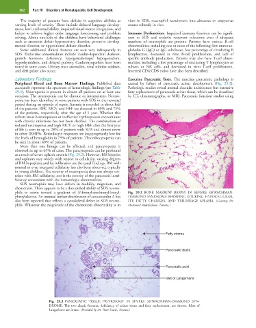
Peripheral Blood and Bone Marrow Findings. Published data caused by failure of pancreatic acinar development (Fig. 29.3).
accurately represent the spectrum of hematologic findings (see Table Pathologic studies reveal normal ductular architecture but extensive
29.4). Neutropenia is present in almost all patients on at least one fatty replacement of pancreatic acinar tissue, which can be visualized
occasion. The neutropenia can be chronic or intermittent. Neutro- by CT, ultrasonography, or MRI. Pancreatic function studies using
penia has been identified in some patients with SDS in the neonatal
period during an episode of sepsis. Anemia is recorded in about half
of the patients. RBC MCV and HbF are elevated in 60% and 75%
of the patients, respectively, after the age of 1 year. Whether this
reflects stress hematopoiesis or ineffective erythropoiesis concomitant
with chronic infections has not been clarified. The combination of
isolated neutropenia and high MCV or high HbF after the first year
of life is seen in up to 28% of patients with SDS and almost never
in other IBMFSs. Reticulocyte responses are inappropriately low for
the levels of hemoglobin in 75% of patients. Thrombocytopenia can
be seen in about 40% of patients.
More than one lineage can be affected, and pancytopenia is
observed in up to 65% of cases. The pancytopenia can be profound
as a result of severe aplastic anemia (Fig. 29.2). However, BM biopsies
and aspirates vary widely with respect to cellularity; varying degrees
of BM hypoplasia and fat infiltration are the usual findings. BM with
normal or even increased cellularity has also been observed, typically
in young children. The severity of neutropenia does not always cor-
relate with BM cellularity, nor is the severity of the pancreatic insuf-
ficiency concordant with the hematologic abnormalities.
SDS neutrophils may have defects in mobility, migration, and
chemotaxis. There appears to be a diminished ability of SDS neutro-
phils to orient toward a gradient of N-formyl-methionyl-leucyl- Fig. 29.2 BONE MARROW BIOPSY IN SEVERE SHWACHMAN-
phenylalanine. An unusual surface distribution of concanavalin A has DIAMOND SYNDROME SHOWING STRIKING HYPOCELLULAR-
also been reported that reflects a cytoskeletal defect in SDS neutro- ITY, FATTY CHANGES, AND TRILINEAGE APLASIA. (Courtesy Dr.
phils. Whatever the magnitude of the chemotaxis abnormality is in Mohamed Abdelhaleem, Toronto.)
Fatty stroma
Pancreatic ducts
Pancreatic acini
Islet of Langerhans
Fig. 29.3 PANCREATIC TISSUE PATHOLOGY IN SEVERE SHWACHMAN-DIAMOND SYN-
DROME. The two classic features, deficiency of acinar tissue and fatty replacement, are shown. Islets of
Langerhans are intact. (Provided by Dr. Peter Durie, Toronto.)