
Page 714 - Hematology_ Basic Principles and Practice ( PDFDrive )
P. 714
606 Part V Red Blood Cells
There is an increased risk for venous thrombosis with an approxi- Sickle Cell–β-Thalassemia
mately twofold increase in risk and sickle trait explaining 7% of
29
thrombotic episodes in African Americans. Armed forces recruits in The gene frequency of β-thalassemia among African Americans is
8
basic training with the sickle cell trait have a substantially increased, 0.004, one-tenth that of the sickle cell gene, and hence there
age-dependent risk of exercise-related sudden death. 30 is one-tenth the prevalence of compound heterozygous sickle cell–β-
Despite the known complications, past experiences with discrimi- thalassemia in this population. Sickle cell–β-thalassemia is divided
+
nation in the employment market and health insurance industry into sickle cell–β -thalassemia and sickle cell–β°-thalassemia, which
provide reminders that the rare clinical events in sickle cell trait have, respectively, reduced or no amounts of Hb A present. Most
+
provide no real justification for regarding it as anything but a benign β-thalassemia mutations among African Americans result in β -
+
31
carrier condition. Newborn screening programs detect a large thalassemia. Sickle cell–β -thalassemia is subclassified according to
number of infants with sickle cell trait; for these parents, genetic the percentage of Hb A present: type I has 3% to 5%, type II has
counseling is essential. Parents should understand that their child has 8% to 14%, and type III has 18% to 25%. Eighty percent of African
a benign hereditary condition with some risks as above but that there American α-thalassemia mutations are attributable to the promoter
is a risk for a subsequent child to be born with SCD. region mutations (−88 [C to T] and −29[A to G]) that result in
In individuals who appear to have sickle cell trait but are symp- a type III phenotype. Compound heterozygous sickle cell–β°-
tomatic, the laboratory diagnosis must be verified. Hemoglobins thalassemia occurs infrequently.
other than S that polymerize may account for reports of “sickle cell In sickle cell–β-thalassemia, the RBCs are hypochromic and
trait” associated with clinical problems. Examples are heterozygous microcytic. The ISCs present on the peripheral blood smear are more
+
Hb S Antilles and Hb Quebec-CHORI. In the latter case, the Hb numerous in sickle cell–β°-thalassemia than in sickle cell–β -
variant was distinguished from Hb A using mass spectroscopy. thalassemia. The hematologic and clinical severity is a function of the
amount of Hb A inherited (Table 42.10).
Additional mitigating influences in sickle cell–β-thalassemia are
Hb SC Disease elevated levels of Hb A 2 and, in sickle cell–β -thalassemia, levels of
+
Hb A up to 30%. These affect both the solubility and polymerization
6
The gene for Hb C (α 2 β 2 Glu→Lys) is approximately one-fourth as of Hb S. Hb F is a more active inhibitor of polymerization than Hb
8
frequent among African Americans as the sickle cell gene. Although A, as shown by Hb S solutions with 15% to 30% Hb A (resembling
+
oxygenated Hb C forms crystals, Hb C does not participate in sickle cell–β -thalassemia) having delay times 10–100 times longer
polymerization with deoxy-Hb S. However, Hb C sustains potassium than pure Hb S solutions, and Hb S solutions with 20% to 30% Hb
chloride cotransport and RBC dehydration, raising the intraerythro- F (resembling Hb S–HPFH) having delay times 1000–1,000,000
cytic concentration of Hb S to levels that support polymerization, times longer. A further influence mitigating the polymerization,
sickling, and clinical symptoms. As a result of a longer circulatory sickling, and clinical aspects of sickle cell–β-thalassemia is the reduced
survival of Hb SC RBCs compared with Hb SS cells (i.e., 27 versus MCHC, which retards Hb S polymerization. Hematologic values for
130
17 days), the degree of anemia and reticulocytosis is frequently sickle cell anemia, the sickle cell–β-thalassemias, and Hb S–HPFH
mild: 75% of the patients have a milder level of anemia (hematocrit are found in Table 42.10.
level >28%) than is usually seen in sickle cell anemia. The predomi-
nant RBC abnormality on the peripheral smear is an abundance of
target cells; folded (“pita bread”) cells, ISCs, “billiard ball” cells, and Sickle Cell–Hb Lepore Diseaseβ
crystal-containing cells may also be seen.
Splenomegaly may be the only physical finding, and the frequency The Hb Lepore gene is a crossover fusion product of the δ- and
of acute painful episodes is approximately half that in Hb SS disease, β-globin genes, the product of which, in the case of Hb Lepore
16
with a life expectancy two decades longer. Nonetheless, significant Boston, has the same alkaline electrophoretic mobility as Hb S.
morbidity can occur. The incidence of fatal bacterial infection is less Therefore patients with the Hb Lepore trait can appear to have sickle
than in sickle cell anemia, but there is still an increased risk of S. cell trait but with only 12% Hb S from thalassemic expression of the
pneumoniae and H. influenzae infection. Osteonecrosis occurs in abnormal fusion gene. Again, because of the electrophoretic similarity
32
approximately 15% of patients. There is a higher incidence of with Hb S, compound heterozygous Hb S–Hb Lepore Boston
peripheral retinopathy in Hb SC disease than in sickle cell anemia. resembles sickle cell anemia or sickle cell–β°-thalassemia electropho-
Coexistent α-thalassemia reduces risk of chronic organ complica- retically but clinically have less severe anemia, resembling that of
+
32
tions. There is an association between renal medullary carcinoma sickle cell–β -thalassemia. The diagnosis is also suggested by the low
and Hb SC disease. to low-normal Hb A 2 levels that result from the incapacitation of one
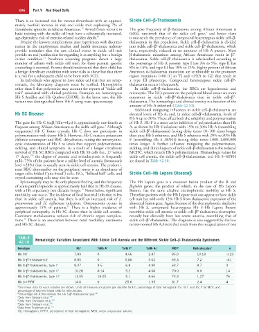
TABLE Hematologic Variables Associated With Sickle Cell Anemia and the Different Sickle Cell–β-Thalassemia Syndromes
42.10
Genotype Hb a %Hb A b %Hb F b %Hb A 2 a MCV a Reticulocytes a n
Hb SS c 7.83 0 4.56 2.87 85.9 10.18 ≈123
Hb S–β°-thalassemia c 8.85 0 5.86 5.02 69.3 7.2 ≈41
Hb S–β -thalassemia, type I d 8.37 3-5 6.8 4.90 63.7 9.7 3
+
Hb S–β -thalassemia, type II d 10.28 8-14 5.2 4.68 70.0 6.6 14
+
Hb S–β -thalassemia, type III e 11.55 18-25 5.1 4.66 73.3 1.27 76
+
Hb S-HPFH f 14.6 0 25.8 1.95 81.7 2.4 4
a The mean data for each variable are shown. Units of measure are grams per deciliter for Hb, percentage of total hemoglobin for Hb F and A2, fl for MCV, and
percentage of total red blood cells for reticulocytes.
+
b Percentage Hb A that defines the Hb S-β -thalassemia type. 247
c Data from Serjeant et al. 249
d Data from Christakis et al. 248
e Data from Serjeant et al. 250
f Data from Friedman et al. 251
Hb, Hemoglobin; HPFH, persistence of fetal hemoglobin; MCV, mean corpuscular volume.