
Page 794 - Hematology_ Basic Principles and Practice ( PDFDrive )
P. 794
680 Part VI Non-Malignant Leukocytes
and as an immune adjuvant in patients with autoimmune disorders Connective Tissue Disorder
and colorectal cancer.
Severe neutropenia is increasingly recognized in patients treated Connective tissue disorders, such as SLE and RA, have been associ-
with rituximab. This neutropenia is unusual for its late onset, gener- ated with monocytosis in the context of chronic inflammation.
ally about 3 months after the last rituximab dose. It occurs with
underlying autoimmune disorders, B-cell malignancies, and stem cell
transplants—basically in all situations in which rituximab is used— Hematopoietic Malignancies
often occurring while the underlying illness is in complete remission.
Severe neutropenia has been reported in about 5% of rituximab- Monocytosis is common with MDS. CMML is defined as persistent
3
treated patients, much higher in some series. Most patients recover peripheral blood monocytosis greater than 1000/mm , absent Phila-
quickly, usually after G-CSF therapy, but there have been protracted delphia chromosome, and evidence of dysplasia in one or more
cases. The authors and others have reported a high risk of relapse hematopoietic cell lineages. Juvenile myelomonocytic leukemia, a
(100% in our series) if rituximab is reinitiated. The mechanism is disease of children that shares pathologic features with CMML,
not definitively established, but an intriguing report implicates imbal- results from defective RAS signaling. Acute myeloid leukemias
anced recovery of B-cell clones with a deficiency of stromal-derived (AMLs) involving the monocyte line (acute myelomonocytic and
factor 1. acute monoblastic leukemias) may release substantial amounts of
lysozyme (muramidase), which is toxic to renal tubules. Serum
lysozyme was used to aid in the diagnosis of these leukemias. Mono-
MONOCYTOSIS cytosis can result from other myeloid leukemias, MPNs, and lym-
phomas, particularly Hodgkin disease.
Monocytosis is extremely nonspecific, usually not requiring investiga-
tion per se. Monocytosis has been defined as a sustained absolute
3
3
increase in monocyte count greater than 800/mm to 1000/mm MONOCYTOPENIA
(Table 48.3). Transient monocytosis, relative or absolute, is common
with recovery from myelosuppression, such as after chemotherapy. Monocytopenia frequently accompanies granulocytopenia with che-
Relative or absolute monocytosis may occur in other myelosuppressed motherapy, aplastic anemia, or other BM-suppressive insults. Isolated
states, such as aplastic anemia. This is a favorable factor associated or disproportionate monocytopenia is rarely recognized, but observa-
with a lower risk of neutropenic infection, perhaps because some tions suggest that this can have serious clinical sequelae when it
phagocytic capacity is maintained. On the other hand, monocytosis occurs. In hairy cell leukemia and monoMAC syndrome (GATA2
is also very common with hematologic malignancies. In a patient with deficiency), there is a clear association with severe opportunistic
unexplained cytopenia, monocytosis can be an important clue to an infections, particularly those normally engendering a granulomatous
underlying MDS. response.
Infectious Diseases Hairy Cell Leukemia
Mycobacterial infection is a common cause of monocytosis world- This disorder, considered in detail elsewhere, classically presents with
wide, related to its propensity for intracellular infection and tissue pancytopenia and splenomegaly, often in middle-aged men. The
granuloma formation. Brucellosis and subacute bacterial endocarditis WBC count is sometimes high because of hairy cell proliferation, but
have also been associated with monocytosis. Certain viral infections, monocytes are invariably severely depressed. (Monocytopenia is not
including influenza, varicella-zoster, CMV, and dengue, have been seen with “variant hairy cell leukemia.”) Neutropenia only partly
reported to cause monocytosis. explains the very high infectious morbidity and mortality. An extraor-
dinary incidence of opportunistic granulomatous infections used to
be encountered, including atypical mycobacteria, tuberculosis, histo-
plasmosis, and other fungi. These are linked to monocytopenia, with
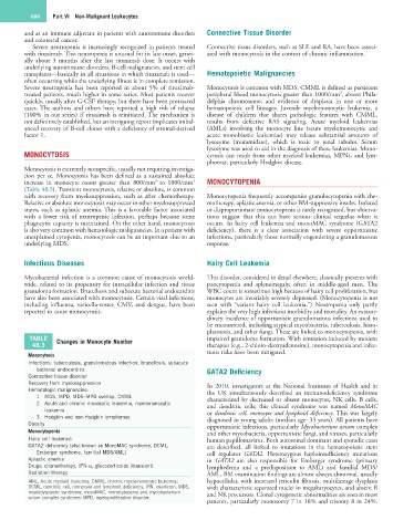
TABLE Changes in Monocyte Number impaired granuloma formation. With remission induced by modern
48.3 therapies (e.g., 2-chloro-deoxyadenosine), monocytopenia and infec-
tious risks have been mitigated.
Monocytosis
Infections: tuberculosis, granulomatous infection, brucellosis, subacute
bacterial endocarditis GATA2 Deficiency
Connective tissue disorder
Recovery from myelosuppression In 2010, investigators at the National Institutes of Health and in
Hematologic malignancies: the UK simultaneously described an immunodeficiency syndrome
1. MDS, MPD, MDS–MPD overlap, CMML characterized by decreased or absent monocytes, NK cells, B cells,
2. Acute and chronic monocytic leukemia, myelomonocytic and dendritic cells; this clinical syndrome was named MonoMAC
leukemia or dendritic cell, monocyte and lymphoid deficiency. This was largely
3. Hodgkin and non-Hodgkin lymphomas diagnosed in young adults (median age: 33 years). All patients have
Obesity opportunistic infections, particularly Mycobacterium avium complex
Monocytopenia and other mycobacteria, opportunistic fungi, and viruses, particularly
Hairy cell leukemia human papillomavirus. Both autosomal dominant and sporadic cases
GATA2 deficiency (also known as MonoMAC syndrome, DCML, are described, all linked to mutations in the hematopoietic stem
Emberger syndrome, familial MDS/AML) cell regulator GATA2. Heterozygous haploinsufficiency mutations
Aplastic anemia in GATA2 are also responsible for Emberger syndrome (primary
Drugs: chemotherapy, IFN-α, glucocorticoids (transient) lymphedema and a predisposition to AML) and familial MDS/
Radiation therapy AML. BM examination findings are almost always abnormal, usually
AML, Acute myeloid leukemia; CMML, chronic myelomonocytic leukemia; hypocellular, with increased reticulin fibrosis, multilineage dysplasia
DCML, dendritic cell, monocyte and lymphoid deficiency; IFN, interferon; MDS, with characteristic separated nuclei in megakaryocytes, and absent B
myelodysplastic syndrome; monoMAC, monocytopenia and mycobacterium and NK precursors. Clonal cytogenetic abnormalities are seen in most
avium complex syndrome; MPD, myeloproliferative disorder.
patients, particularly monosomy 7 in 16% and trisomy 8 in 24%.