
Page 276 - Williams Hematology ( PDFDrive )
P. 276
250 Part IV: Molecular and Cellular Hematology Chapter 17: Signal Transduction Pathways 251
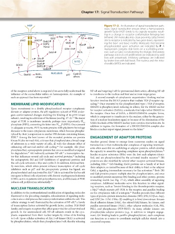
HGF Figure 17–2. An illustration of signal transduction path-
ways. Signal transduction ensues when a hematopoietic
growth factor (HGF) binds to its cognate receptor, result-
PIP2 Ras Raf1 PIP3 ing in a change in receptor conformation bringing two
SOS Akt PDK tethered JAK molecules into close proximity (attachment
JAK2 JAK2-P Gab2-P site to receptor is indicated by two green boxes, represent-
SHP2 MEK1-P Bad-P ing the box1 and box2 motifs). Molecules that become
?Grb2 GSK-P phosphorylated upon activation are indicated by P. A
ERK1/2-P multiprotein complex that forms on a scaffolding mole-
Grb2-P BclXL cule, such as Gab2, is indicated by the triangle. Stimulatory
pathways (vis-à-vis cell proliferation) are indicated by solid
Y Y-P Shc-P lines with arrowheads. Inhibitory pathways are indicated
Stat3-P MITO by broken lines with ball heads. The nucleus and the mito-
Stat5-P chondria (MITO) are indicated.
SOCS
SHP2-P p85
Gab2-P
StatRE BclXL, NUCLEUS p27 CyclinD,
others others
of the receptors noted above is required if we are to fully understand the NF-κB and targeting I-κB for proteasomal destruction, allowing NF-κB
influence of the extracellular milieu on hematopoiesis. An example of to translocate to the nucleus and bind and activate target genes.
such an approach has been reported. 63 A second example of cytoplasmic sequestration blocking nuclear
function involves the SMAD proteins that mediate TGF-β receptor sig-
MEMBRANE LIPID MODIFICATION naling. Once recruited to the phosphorylated type I TGF-β receptor,
36
Upon recruitment to a doubly phosphorylated receptor cytoplasmic SMAD2 is phosphorylated, reducing its affinity for the SMAD anchor
for receptor activation (SARA), a molecule that helps tether SMAD2 to
domain or adapter protein, the p85 regulatory subunit of PI3K under- the receptor. Once free of SARA, a SMAD2/SMAD4 complex forms,
goes conformational changes enabling the binding of its p110 kinase which is competent to translocate to the nucleus, either by the genera-
subunit, resulting in activation of the kinase (see Fig. 17–2). The major tion of a nuclear localization signal or because of the elimination of the
64
target of PI3K is membrane inositols, perhaps most importantly PI SARA blockade of the SMAD2 nuclear pore complex interaction site. In
4,5
phosphate (PIP2), converting the latter into PI 3,4,5 P (PIP3). Once present addition to ingress, the formation of a SMAD2/SMAD4 complex also
in adequate amounts, PIP3 recruits proteins with pleckstrin homology blocks a nuclear export signal present on the latter. 71
domains to the inner cytoplasmic membrane, which become phospho-
rylated by their juxtaposition to another PH domain containing kinase,
PDK. Among the best known of the recruited proteins are protein ENGAGEMENT OF ADAPTOR PROTEINS
65
kinase B (also termed Akt), a kinase that phosphorylates a broad range Another general theme to emerge from numerous studies on signal
of substrates in a wide variety of cells, all with the ultimate effect of transduction is that multimolecular complexes of signaling intermedi-
enhancing cell survival and/or cell cycling. For example, Akt phos- aries often assemble on scaffolding or adaptor proteins, which develop
66
phorylates Bad, a proapoptotic protein that once so modified is targeted the capacity to assemble signaling complexes upon phosphorylation.
72
for degradation. Akt indirectly activates NF-κB, a transcription fac- Insulin receptor substrates (IRSs) were the first such adaptors identi-
67
67
tor that influences several cell-cycle and survival proteins, including fied, and are phosphorylated by the activated insulin receptor. IRS
68
73
the antiapoptotic Bcl and IAP (inhibitors of apoptosis) proteins and proteins are also modified by several other receptor-activated kinases,
the cell-cycle activators c-Myc and cyclin D. In addition, forkhead fam- including JAKs. Grb-binding (Gab) proteins are a family of at least
54
ily members, which when present enhance transcription of cell-cycle three adapters, so named because of their ability to bind to the adaptor
inhibitors such as p27 and the proapoptotic protein Fas ligand, are Grb2, a signaling intermediate necessary for Ras activation. Both IRS
74
phosphorylated and inactivated by Akt. Akt is activated by the bcr-abl and Gab proteins present multiple sites for phosphorylation, and once
69
oncogene in blood cells of patients with chronic myelogenous leukemia so modified present numerous SH2-binding and other protein–protein
(CML), as it is in patients with polycythemia vera, and blockade of PI3K interacting motifs (see Fig. 17–2), which allow assembly of signaling
reduces cell proliferation substantially. 70 complexes. Additional molecules serve this function in other signal-
NUCLEAR TRANSLOCATION ing receptors, such as Tensin2 binding to the thrombopoietin receptor,
c-Mpl, which recruits p85 PI3K to the receptor, and paxillin binding
63
In addition to the posttranslational modification of signaling molecules on the cytoplasmic tails of α-integrin. Paxillin presents four different
48
illustrated in the preceding examples, relocalization of signaling mole- types of protein–protein interaction domains (SH3, SH2, LD [Leu-Asp],
cules is also a vital process that conveys information within the cell. This and LIM [lin-11/Isl-1/Mec-3]) enabling it to bind downstream kinases
cellular strategy is well illustrated by the activation of NF-κB, a family (focal adhesion kinase [FAK], the related Pyk2 kinase, Src kinase, and
68
of transcription factors activated by growth factor, nuclear, TGF/BMP paxillin-associated kinase [PAK]), other adaptor molecules (Crk, PIX,
family, and integrin receptors that affect genes vital for cell survival and PKL), and phosphatases (PTP-PEST). As many cellular kinases can
growth. In the unstimulated cell, NF-κB subunits reside in the cyto- phosphorylate adaptor proteins (e.g., in addition to integrin engage-
plasm, sequestered from their nuclear targets by virtue of its binding ment, GH binding leads to paxillin phosphorylation), such complexes
to I-κB. Upon cellular activation of Akt, I-κB kinase (IKK) is activated can function as a nexus to coordinate multiple cellular stimuli into a
by phosphorylation, which then phosphorylates I-κB, thereby releasing concerted response.
Kaushansky_chapter 17_p0247-0256.indd 251 9/17/15 5:45 PM