
Page 283 - Williams Hematology ( PDFDrive )
P. 283
258 Part IV: Molecular and Cellular Hematology Chapter 18: Hematopoietic Stem Cells, Progenitors, and Cytokines 259
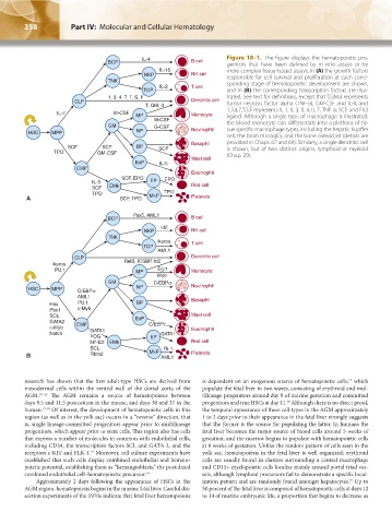
Figure 18–1. The figure displays the hematopoietic pro-
genitors that have been defined by in vitro assays or by
more complex tissue-based assays. In (A) the growth factors
responsible for cell survival and proliferation at each corre-
sponding stage of hematopoietic development are shown,
and in (B) the corresponding transcription factors are illus-
trated. See text for definitions, except that T,GM,4 represents
tumor necrosis factor alpha (TNF-α), GM-CSF and IL-4, and
1,3,4,7,T,S,F represents IL-1, IL-3, IL-4, IL-7, TNF-α, SCF, and Flt3
ligand. Although a single type of macrophage is illustrated,
the blood monocyte can differentiate into a plethora of tis-
sue specific macrophage types, including the hepatic Kupffer
cell, the brain microglia, and the bone osteoclast (details are
provided in Chaps. 67 and 69). Similarly, a single dendritic cell
is shown, but of two distinct origins, lymphoid or myeloid
(Chap. 20).
research has shown that the first adult-type HSCs are derived from is dependent on an exogenous source of hematopoietic cells, which
17
mesodermal cells within the ventral wall of the dorsal aorta of the populate the fetal liver in two waves, consisting of erythroid and mul-
AGM. 10–12 The AGM remains a source of hematopoiesis between tilineage progenitors around day 9 of murine gestation and committed
days 9.5 and 11.5 postcoitum in the mouse, and days 30 and 37 in the progenitors and true HSCs at day 11. Although there is no direct proof,
18
human. 13,14 Of interest, the development of hematopoietic cells in this the temporal appearance of these cell types in the AGM approximately
region (as well as in the yolk sac) occurs in a “reverse” direction, that 1 to 2 days prior to their appearance in the fetal liver strongly suggests
is, single lineage-committed progenitors appear prior to multilineage that the former is the source for populating the latter. In humans the
progenitors, which appear prior to stem cells. This region also has cells fetal liver becomes the major source of blood cells around 5 weeks of
that express a number of molecules in common with endothelial cells, gestation, and the marrow begins to populate with hematopoietic cells
including CD34, the transcription factors SCL and GATA-2, and the at 8 weeks of gestation. Unlike the random pattern of cells seen in the
receptors c-KIT and FLK-1. Moreover, cell culture experiments have yolk sac, hematopoiesis in the fetal liver is well organized; erythroid
15
established that such cells display combined endothelial and hemato- cells are usually found in clusters surrounding a central macrophage
poietic potential, establishing them as “hemangioblasts,” the postulated and CD15+ myelopoietic cells localize mainly around portal triad ves-
combined endothelial cell–hematopoietic precursor. 16 sels, although lymphoid precursors fail to demonstrate a specific local-
Approximately 2 days following the appearance of HSCs in the ization pattern and are randomly found amongst hepatocytes. Up to
19
AGM region, hematopoiesis begins in the murine fetal liver. Careful dis- 50 percent of the fetal liver is composed of hematopoietic cells at days 12
section experiments of the 1970s indicate that fetal liver hematopoiesis to 14 of murine embryonic life, a proportion that begins to decrease as
Kaushansky_chapter 18_p0257-0278.indd 258 9/19/15 12:05 AM