
Page 288 - Williams Hematology ( PDFDrive )
P. 288
262 Part IV: Molecular and Cellular Hematology Chapter 18: Hematopoietic Stem Cells, Progenitors, and Cytokines 263
Several investigators have shown that the 5′ flanking region of the c-mpl Perivascular
gene contains a functionally important GATA site and that GATA1 Mø Stromal cells
transactivates the gene in hematopoietic cell lines. Because GATA1 CAR cells
134
does not appear in hematopoietic cells until they have lost their repop- Nestin-GFP +
ulating capacity, it is possible that GATA2 fulfills this role in HSCs, LeptinR +
although there is no evidence yet available establishing that this protein Prx1 +
can transactivate the c-mpl GATA site. 135
STEM CELL AGING Mø HSC `SM
While blood cell counts do not change substantially in elderly mam- CXCL 12
mals, a number of alterations are demonstrable in HSCs derived from OM
older individuals. In aged mice, the HSC compartment expands, HSC OM KitL
136
although each HSC has reduced capacity to expand. Upon trans-
plantation, aged marrow HSCs display a myeloid skewing, generating Ob Oc
reduced numbers of T- and B-lymphoid precursors, that along with
thymic involution helps to explain the immune depletion seen in older BONE
adults. Similar findings were reported when aged human stem cells were
137
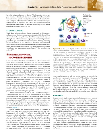
transplanted into immunocompromised mice. This topic has been Figure 18-2. The figure depicts multiple elements of the hemato-
reviewed. 138 poietic microenvironment. The niche has two major regions in the
marrow, supported by osteoblasts or vascular cells. Several cell types
THE HEMATOPOIETIC provide cytokines that maintain osteoblasts (Ob), which, in turn, sup-
port hematopoietic stem cells (HSC) by secreting CXCL12 and other
MICROENVIRONMENT cytokines. Osteoclasts (Oc) are also shown but are of lesser importance
in HSC maintenance, and may inhibit HSC survival/proliferation. Mac-
It has been estimated that the concentration of cells within the mar- rophages that express α smooth muscle actin (αSM) support perivas-
row is 10 /mL; as a result, multiple cell–cell and cell–matrix interac- cular cells, including the CXCL12 abundant reticular (CAR) cells, that, in
9
tions occur. A major advance in experimental hematology has been the turn, provide CXCL12 and SCF (here termed c-kit ligand [KitL]) to HSCs. In
capacity to grow hematopoietic cells in long-term culture. When high addition to paracrine support, direct perivascular cell–stem cell contact,
concentrations of marrow cells are placed in serum-containing cultures, through integrins, also support HSCs. (Reproduced with permission from
a stromal cell layer and extracellular proteinaceous matrix form, and Calvi LM, Link DC: Cellular complexity of the bone marrow hematopoietic
when subsequently recharged with fresh marrow cells, these long-term stem cell niche. Calcif Tissue Int 94(1):112–124, 2014.)
cultures (LTCs) are capable of supporting hematopoiesis for months
with simple demidepletion and replacement of culture medium. It is present on hematopoietic cells and counterreceptors on stromal cells
assumed that the cell–cell and cell–matrix interactions that develop are also very important for hematopoiesis. 65,72 In addition to bringing
in such cultures more closely resemble those found in vivo, helping to hematopoietic cells into close proximity to cells producing soluble or
explain the longevity of such cultures and their capacity to maintain cell-bound cytokines, and hence raising the local concentration of these
hematopoietic stem and primitive progenitor cells far longer ex vivo growth promoting proteins, integrin engagement leads to intracellular
than do nonstromal cell-containing cultures. The molecular basis for signaling, usually promoting entry into the cell cycle and preventing
the improved hematopoietic environment of LTCs is thought to rely on programmed cell death. Reflecting the vital and sometimes lineage
148
stromal cell surface molecules that promote cell–cell contact, prevent specific roles of the hematopoietic microenvironment, the extracellular
programmed cell death, and regulate growth. matrix and stromal cells reside in a highly organized structure (Chap. 5).
The microenvironmental effects on HSCs have far reaching clin-
ical implications as well; our ability to mobilize marrow stem cells for
transplantation has greatly changed the way we treat hematologic and ANATOMY
other malignancies, and ultimate success in the efforts of experimental Hematopoiesis is highly compartmentalized within areas of red marrow,
hematologists to expand HSCs ex vivo with cocktails of cytokines and with erythropoiesis occurring in clusters surrounding a central macro-
150
149
stromal cells for applications in gene therapy and regenerative medi- phage, granulocyte development associated with stromal cells, and
151
cine will undoubtedly derive only from a thorough understanding of megakaryopoiesis occurring adjacent to the endothelial sinusoidal cells.
the molecular bases for the interaction of HSCs with their microenvi- In the adult marrow, the specialized niche in which HSCs develop into
ronment (Fig. 18–2). differentiated progeny has been termed the hematon by Peault, a structure
Marrow stromal cells influence hematopoiesis in a number of that includes Str01+ mesenchymal cells, desmin-positive perivascular
ways, by producing several cytokines that positively or negatively affect lipocytes, Flk1+ endothelial cells, macrophages, and hematopoietic pro-
152
hematopoietic cell growth, 139–142 including some, like SCF, that are genitors. From these structures can be derived all lineages of committed
expressed on their cell surfaces, resulting in enhanced biologic activ- colony-forming cells (e.g., CFU-GM and BFU-E) and primitive cells that
143
ity. Stromal cells are the origin of a number of extracellular matrix pro- score positive in CAFC assays, LTC-IC, and high proliferative potential
teins that either directly affect hematopoietic cells, or do so indirectly by colony-forming cell assays (Chap. 5).
binding growth factors and presenting them in a functional context. One consequence (or perhaps cause) of this anatomical arrange-
144
They also bear the Jagged/Delta family ligands that stimulate Notch ment is that the stem cell microenvironment is quite hypoxia. It is esti-
proteins to undergo cleavage and translocation into the nucleus, events mated that the O level of the stem cell niche is approximately 5 percent.
2
that are critical mediators of cell fate decision making, 145,146 including The HSC response to hypoxia is discussed in “Metabolic Characteris-
for hematopoietic cells. Cell–cell interactions mediated by integrins tics” above.
147
Kaushansky_chapter 18_p0257-0278.indd 263 9/19/15 12:05 AM