
Page 707 - Williams Hematology ( PDFDrive )
P. 707
682 Part VI: The Erythrocyte Chapter 46: Erythrocyte Membrane Disorders 683
Other Neuroacanthocytosis Syndromes clinical phenotypes and also resemble Huntington disease, which ren-
The HDL2 disorder is caused by expanded CGT/CAG trinucleotide ders the clinical diagnosis difficult. Identification of the underlying gene
repeat mutations in the junctophilin-3 gene, which encodes a protein defects and the availability of molecular tests have markedly improved
involved in junctional membrane structures and calcium regulation. the diagnostic accuracy. This also provides insight into the underlying
167
The disease is autosomal dominant and presents with late-onset cho- pathogenesis and suggests that the affected proteins, which are all linked
rea, parkinsonism, and progressive cognitive defects. Acanthocytes are to membrane structure, may participate in a common pathway that ulti-
present in some patients. In one unusual kindred autosomal dominant mately causes degeneration of the basal ganglia.
inheritance of chorea-acanthocytosis with polyglutamine neuronal
inclusions was described in association with HDL2. Proteolysis of
band 3 was also noted, which could contribute to the altered red cell HEREDITARY STOMATOCYTOSIS SYNDROMES
morphology. 170,173 The intracellular concentration of the monovalent cations, Na and K ,
+
+
Acanthocytes have been noted in some patients with PKAN (for- contribute to erythrocyte volume homeostasis. A net increase in these
merly known as Hallervorden-Spatz syndrome) with features of dys- cations causes water to enter the cells resulting in overhydrated cells or
tonia, dysarthria, and rigidity in childhood, and in HARP syndrome stomatocytes, whereas a net loss dehydrates the cells and forms xero-
(hypobetalipoproteinemia, acanthocytosis, retinitis pigmentosa and cytes. Disorders of red cell cation permeability are very rare conditions
pallidal degeneration). Both conditions are caused by mutations in pan- that are inherited in an autosomal dominant fashion with marked clini-
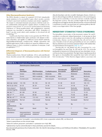
tothenate kinase 2, which is involved in synthesis of coenzyme A and cal and biochemical heterogeneity (Table 46–4). 175
phospholipids. 167,170,174 Stomatocytes are cup-shaped red cells characterized by a cen-
tral hemoglobin-free area (Figs. 46–10E and 46–11D). The molecular
Differential Diagnosis of Neuroacanthocytosis with Normal mechanism of stomatocyte formation has not been elucidated, but sev-
Lipoproteins eral theories have been postulated. The lipid bilayer hypothesis predicts
Chorea-acanthocytosis, McLeod syndrome, HDL2, and pantothenate that agents or abnormalities that expand the inner leaflet will tend to
kinase disorders present with overlapping neurologic symptoms and form stomatocytes. Other theories relegate lipids to a secondary role
176
TABLE 46–4. Heterogeneity of the Hereditary Stomatocytosis Syndromes
Stomatocytosis (Hydrocytosis) Intermediate Syndromes
Xerocytosis with
Severe Stomatocytic High Phosphati-
Hemolysis Mild Hemolysis Cryohydrocytosis Xerocytosis dylcholine Xerocytosis
Hemolysis Severe Mild–moderate Moderate Mild Moderate Moderate
Anemia Severe Mild–moderate Mild–moderate None Mild Moderate
Blood film Stomatocytes Stomatocytes Stomatocytes Stomatocytes Targets Targets,
echinocytes
MCV (80–100 fL) * 110–150 95–130 90–105 91–98 84–92 100–110
MCHC (32–36%) 24–30 26–29 34–40 33–39 34–38 34–38
Unincubated Markedly Increased Normal Decreased Markedly Markedly
osmotic fragility increased decreased decreased
RBC Na +5–12† 60–100 30–60 40–50 10–20 10–15 10–20
RBC K +90–103 20–55 40–85 55–65 75–85 75–90 60–80
RBC Na +K +95–110 110–140 115–145 100–105 87–103 93–99 75–90
+
Phosphatidylcho- Normal ± Increased Normal Normal Increased Normal
line content
Cold No No Yes No No ?
autohemolysis
Effect of Good Good Fair ? ? ? Poor
splenectomy ‡
Inheritance Autosomal domi- Autosomal Autosomal Autosomal Autosomal Autosomal
nant?, autosomal dominant dominant dominant dominant dominant
recessive
MCHC, mean corpuscular hemoglobin concentration; MCV, mean corpuscular volume; RBC, red blood cell.
* Values in parentheses are the normal range.
† Values for sodium, potassium, and sodium + potassium are mEq/L RBC.
‡ Splenectomy may be contraindicated in these syndromes; see text for details.
Reproduced with permission from Nathan DG, Orkin SH, Oski FA: Hematology of Infancy and Childhood, 5th edition. Philadelphia, PA: Saunders/
Elsevier; 1998.
Kaushansky_chapter 46_p0661-0688.indd 682 9/17/15 6:42 PM