
Page 702 - Williams Hematology ( PDFDrive )
P. 702
676 Part VI: The Erythrocyte Chapter 46: Erythrocyte Membrane Disorders 677
HEREDITARY ELLIPTOCYTOSIS AND rearrangement of the skeleton is reduced. Disruption of the dynamic
PYROPOIKILOCYTOSIS dissociation and reassociation of spectrin tetramers causes mechanical
instability of the membrane, which precludes the recovery of the normal
Definition and History biconcave disk shape of the cell after prolonged and repeated unidirec-
HE is characterized by the presence of elliptical or oval erythrocytes on tional axial distortion in the microcirculation. HE reticulocytes have
127
the blood films of affected individuals (Figs. 46–10D and 46–11F.). In a normal shape when released into the circulation but the mature red
1904, Dresbach, a physiologist at Ohio State University in Columbus, cells become progressively more elliptical as they age and ultimately the
Ohio, published the first description of elliptical red blood cells in one of abnormal shape becomes permanent. 13,122 As the severity of the defect
his students, noticed during a laboratory exercise in which the students increases, poikilocytes are formed and the cells become prone to frag-
were examining their own blood. The report elicited controversy mentation. HPP patients exhibit a combination of horizontal (impaired
119
because the student died soon thereafter, leading to speculation that he spectrin tetramer formation) and vertical (spectrin deficiency) defects,
had actually suffered from pernicious anemia. The demonstration of with the latter causing microspherocytes and exacerbating the hemo-
elliptocytosis in three generations of one family established the hered- lytic anemia. 128,129
itary nature of this disorder. A related disorder, HPP is a rare disease
120
first described in 1975 in children with severe neonatal anemia with Red Cell Membrane Protein Defects
abnormal poikilocytic red cell morphology reminiscent of that seen in Spectrin Mutations that affect spectrin heterodimer self-association
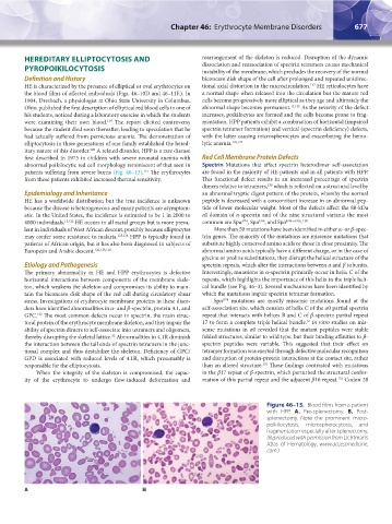
patients suffering from severe burns (Fig. 46–13). The erythrocytes are found in the majority of HE patients and in all patients with HPP.
121
from these patients exhibited increased thermal sensitivity. This functional defect results in an increased percentage of spectrin
dimers relative to tetramers, which is reflected on a structural level by
130
Epidemiology and Inheritance an abnormal tryptic digest pattern of the protein, whereby the normal
HE has a worldwide distribution but the true incidence is unknown peptide is decreased with a concomitant increase in an abnormal pep-
because the disease is heterogeneous and many patients are asymptom- tide of lower molecular weight. Most of the defects affect the 80-kDa
atic. In the United States, the incidence is estimated to be 1 in 2000 to αI domain of α-spectrin and of the nine structural variants the most
.
I/65
4000 individuals. 13,122 HE occurs in all racial groups but is more preva- common are Spα , Spα , and Spα I/46 or 50a 128
I/74
lent in individuals of West African descent, possibly because elliptocytes More than 50 mutations have been identified in either α- or β-spec-
may confer some resistance to malaria. 123,124 HPP is typically found in trin genes. The majority of the mutations are missense mutations that
patients of African origin, but it has also been diagnosed in subjects of substitute highly conserved amino acids or those in close proximity. The
European and Arabic descent. 122,125,126 abnormal amino acids typically have a different charge, or in the case of
glycine or proline substitutions, they disrupt the helical structure of the
Etiology and Pathogenesis spectrin repeats, which alter the interactions between α and β subunits.
The primary abnormality in HE and HPP erythrocytes is defective Interestingly, mutations in α-spectrin primarily occur in helix C of the
horizontal interactions between components of the membrane skele- repeats, which highlights the importance of this helix in the triple heli-
ton, which weakens the skeleton and compromises its ability to main- cal bundle (see Fig. 46–3). Several mechanisms have been identified by
tain the biconcave disk shape of the red cell during circulatory shear which the mutations impair spectrin tetramer formation.
stress. Investigations of erythrocyte membrane proteins in these disor- Spα mutations are mostly missense mutations found at the
I/74
ders have identified abnormalities in α- and β-spectrin, protein 4.1, and self-association site, which consists of helix C of the α0 partial spectrin
GPC. The most common defects occur in spectrin, the main struc- repeat that interacts with helices B and C of β-spectrin partial repeat
122
34
tural protein of the erythrocyte membrane skeleton, and they impair the 17 to form a complete triple helical bundle. In vitro studies on mis-
ability of spectrin dimers to self-associate into tetramers and oligomers, sense mutations in α0 revealed that the mutant peptides were stable
thereby disrupting the skeletal lattice. Abnormalities in 4.1R diminish folded structures, similar to wild type, but their binding affinities to β-
55
the interaction between the tail ends of spectrin tetramers in the junc- spectrin peptides were variable. This suggested that their effect on
tional complex and thus destabilize the skeleton. Deficiency of GPC/ tetramer formation was exerted through defective molecular recognition
GPD is associated with reduced levels of 4.1R, which presumably is and disruption of protein-protein interactions at the contact site, rather
131
responsible for the elliptocytosis. than an altered structure. These findings contrasted with mutations
When the integrity of the skeleton is compromised, the capac- in the β17 repeat of β-spectrin, which perturbed the structural confor-
132
ity of the erythrocyte to undergo flow-induced deformation and mation of this partial repeat and the adjacent β16 repeat. Codon 28
Figure 46–13. Blood films from a patient
with HPP. A. Pre-splenectomy. B. Post-
splenectomy. Note the prominent micro-
poikilocytosis, microspherocytosis, and
fragmentation especially after splenectomy.
(Reproduced with permission from Lichtman’s
Atlas of Hematology, www.accessmedicine.
com.)
A B
Kaushansky_chapter 46_p0661-0688.indd 677 9/17/15 6:42 PM