
Page 168 - Clinical Immunology_ Principles and Practice ( PDFDrive )
P. 168
150 ParT ONE Principles of Immune Response
induction of transcription. The SMAD MH1 motif mediates
sequence-specific DNA binding, whereas the MH2 domain
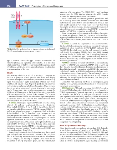
TGF-b RI
receptor contains the transcriptional activation domain.
SMAD2-null mice lack anterior/posterior specification and
fail to develop mesoderm. SMAD3-deficient mice have limb
RII P RSmad malformations and defective immune function. SMAD3-null
P mice exhibit defective TGF-β responses. However, these mice
K K
also display accelerated wound healing compared with normal
mice, which seems to contradict the role of SMAD3 as a positive
regulator of TGF-β in enhancing wound healing.
P Upon recruitment to their cognate activated type I receptor,
Smad4 RSmad R-SMADs are phosphorylated on C-terminal serine residues,
triggering homodimerization of R-SMADs or heterodimerization
P Target genes with another class of SMAD, the common SMAD or C-SMAD
RSmad (see below).
C-SMADs. SMAD4 is the only known C-SMAD in vertebrates.
Smad4 It is thought to function as the central and essential downstream
mediator of other SMADs in all TGF-β/BMP pathways. The
FIG 9.9 SMADs and signaling by transforming growth factor-β
(TGF-β) superfamily receptor serine kinases. SMAD MH2 domain is important for both receptor interaction
and SMAD dimerization. SMAD4 lacks the SSXS element
conserved in the R-SMADs and thus is not phosphorylated,
rendering it unable to bind type I receptors directly. SMAD4-
deficient mice die early in embryogenesis and exhibit severe
type II receptor. In turn, the type I receptor is responsible for defects in gastrulation.
phosphorylating key signaling intermediates. It is not clear I-SMADs. The third subfamily of SMADs is the inhibitory
whether activation of the type I receptor results from enhancement SMADs, or I-SMADs. In mammals, SMAD6 and SMAD7 are
of its kinase activity, the appearance of substrate-binding sites, the I-SMADs. SMAD6 utilizes an alternative mechanism and is
or a combination of the two. thought to function by competing with SMAD4 for binding to
an R-SMAD. Studies with SMAD6-deficient mice indicate a role
SMADs in the development and homeostasis of the cardiovascular system.
The primary substrates activated by the type I receptors are SMAD7 is induced by TGF-β and binds to TGF-β receptors
SMADs, a group of related proteins that have been highly inhibiting the phosphorylation of R-SMADs, thus serving as a
conserved throughout evolution and play a critical role in TGF-β classic feedback inhibitor.
82
signal transduction. Eight mammalian SMADs have been Consistent with their key functions in development, gene
identified. All exhibit a high degree of specificity for conserved targeting of SMAD molecules has been shown to produce severe
motifs in the cytoplasmic tail of type I receptors. These proteins phenotypic abnormalities.
do not contain any previously known structural or enzymatic SMAD pathways. Although a canonical SMAD DNA-binding
motifs. However, they have two homology domains, termed the element (SBE) has been described (AGAC), comparison of the
MH1 and MH2 domains, at the N- and C-termini, respectively, TGF-β responsiveness of synthetic promoters to natural promoters
which are separated by a central linker domain. The extreme has revealed that SMADs can only partially account for the
C-terminus of some SMADs is a critical site of phosphorylation, gene-regulatory effects of TGF-β signaling. SMADs also interact
as described below. SMADs have been subdivided into three with a variety of other transcription factors, transcriptional
classes based on functional distinctions. coactivators, and transcriptional corepressors to regulate transcrip-
R-SMADs. The receptor-regulated SMADs (R-SMADs) directly tion of a select subset of complex promoters in a coordinated
interact with, and are phosphorylated by, the type I receptor. manner. For example, FAST-1 (forkhead activin signal transducer),
These include SMAD1, -2, -3, -5, and -8. SMAD2 and SMAD3 a winged helix forkhead transcription factor, associates with the
are phosphorylated in response to TGF-β, whereas SMAD1, -5, SMAD2/SMAD4 complex. SMADs also bind to c-JUN/c-FOS,
and -8 are primarily activated in response to BMP activation. and AP-1 sites frequently overlap with SBE sites in the naturally
R-SMADs bind to the GS domain of type I receptor and are occurring promoters of TGF-β–responsive genes. SMADs can
phosphorylated at a conserved SSXS motif in their C terminus. also bind ATF2, the vitamin D receptor, and other transcription
The interaction of R-SMADs with TGF-β receptors can also be factors and can recruit the coactivators CBP/p300. Additionally,
regulated by another molecule termed SARA (SMAD anchor the SKI and SNON corepressors can interact with SMADs and
for receptor activation). SARA binds unphosphorylated SMAD2 antagonize TGF-β signaling.
and SMAD3. By virtue of its FYVE domain, SARA can bind
phospholipids and localize SMADs to the plasma membrane, Other TGF-β–Activated Pathways
facilitating receptor binding. Phosphorylation of SMADs also Other signaling pathways are known to mediate TGF-β signals.
permits dissociation from SARA. In particular, a number of members of the MAPK family are
Once phosphorylated, R-SMADs dissociate from the activated activated in response to TGF-β. TGF-β induces ERK, JNK, and
type I receptor and associate with SMAD4 in the cytoplasm. p38 MAPK activity. Inhibition of several of these pathways inhibits
This is followed by nuclear translocation of the heteromeric TGF-β–mediated transcriptional activation. In addition, a MAPK
SMAD complex, the binding to cognate DNA motifs in the family member, TAK1, has been implicated in TGF-β signaling.
promoters of TGF-β–responsive genes, and the concomitant TAK1 is activated in response to ligand binding and has been