
Page 1643 - Hall et al (2015) Principles of Critical Care-McGraw-Hill
P. 1643
1162 PART 10: The Surgical Patient
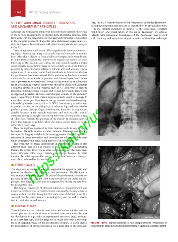
SPECIFIC ABDOMINAL INJURIES—DIAGNOSIS (Fig. 120-6). A free perforation of the first portion of the duodenum pro-
AND MANAGEMENT PRINCIPLES duces pneumoperitoneum and can be identified on an upright chest film.
In the surgical treatment of injuries to the duodenum, complete
Although the nonsurgeon intensivist does not need detailed knowledge mobilization and visualization of the entire duodenum are crucial.
of the surgical management of specific intra-abdominal injuries, some Patients with intramural hematomas of the duodenum may present
familiarity with the diagnostic and management principles to be applied with vomiting and symptoms of gastric outlet obstruction; radiologic
in the surgical treatment of specific intra-abdominal organ injuries is
likely to improve the confidence with which these patients are managed
in the ICU.
Penetrating abdominal injury differs significantly from nonpenetrat-
ing injury. Penetrating injury may result from stab wounds or wounds
https://kat.cr/user/tahir99/
from other sharp objects or from a bullet or shotgun. Stab wounds tend
to be the least serious, in that they involve organs only within the short
trajectory of the weapon, and unless the stab wound impales a major
vessel directly, major hemorrhage is not as likely as in other forms of
penetrating or blunt abdominal injury. Patients with stab wounds require
exploration of the wound under local anesthesia to determine whether
the peritoneum has been violated. If the peritoneum has been violated,
a decision has to be made to proceed with formal laparotomy unless
one is prepared to use peritoneal lavage or ultrasound as an adjunctive
test in determining whether laparotomy should be conducted. Although
a selective approach using imaging such as CT and MRI to identify
tangential nonpenetrating wounds that would not require laparotomy
is suggested, generally, all bullet and shotgun wounds to the abdomen
require laparotomy. These missile injuries usually result in damage to
more than one organ. Since kinetic energy transfer is affected most sig-
nificantly by missile velocity (K = ½ MV ), low-velocity missiles tend
2
to produce limited surrounding injury, whereas high-velocity missiles
produce greater damage. Organ involvement, therefore, is very unpre-
dictable because of the variable trajectory and wide variable area of
dissipated energy. A straight line joining the points of entry and exit usu-
ally does not represent the pathway of the missile. In shotgun injuries,
much less damage is inflicted when the injury occurs from far range
rather than close range.
The crushing force produced by blunt injuries results in very irregular
lacerations. Multiple injuries are also common. Diagnosis and therapy
are more challenging and should be more aggressive. Hemorrhage, devi-
talization of tissue, morbidity, and mortality are all increased in blunt
injury compared with penetrating injuries of the abdomen.
The frequency of organ involvement in penetrating trauma is also
different from that in blunt trauma to the abdomen. In penetrating
trauma, the organs involved, in order of frequency, are the liver, small
bowel, stomach, colon, major vessels, and retroperitoneum. In blunt
injuries, the solid organs—the spleen, kidney, and liver—are damaged
most often, followed by the intestines.
■ STOMACH INJURIES
The diagnosis of stomach injury is suggested by epigastric pain and
pain at the shoulder tip if there is free perforation. Usually there is
very minimal hemorrhage, and the patient’s hemodynamic status is not
particularly affected. Upright chest x-ray reveals free air under the dia-
phragm. The diagnosis also may be suggested by bloody aspirate from
the nasogastric tube.
The surgical treatment of stomach injuries is straightforward and
involves débridement of devitalized tissue and usually primary suture or
anastomosis if resection is required for wide areas of devitalization. It is
essential that the entire stomach, including the posterior wall, is visual-
ized to minimize missed injuries.
■ DUODENAL INJURIES
These injuries are seen often in association with other injuries, and the
second portion of the duodenum is involved most commonly. Because
the duodenum is a partially retroperitoneal structure, frank peritoni-
tis is a very late sign, and the diagnosis is made only with a very high
index of suspicion based on the mechanism of injury. A useful sign is FIGURE 120-6. Ruptured duodenum. A. Plain radiograph showing retroperitoneal air
the identification of retroperitoneal air on a plain film of the abdomen around the right kidney. B. Transected duodenum found at laparotomy on the same patient.
section10.indd 1162 1/20/2015 9:21:11 AM