
Page 1640 - Hall et al (2015) Principles of Critical Care-McGraw-Hill
P. 1640
CHAPTER 120: Torso Trauma 1159
■ BLUNT CARDIAC INJURY suggestive sign is a widening of the mediastinum on the plain chest film
This lesion probably occurs much more commonly than was suspected (Fig. 120-5). Other suggestive signs are the presence of a thoracic bruit
or a discrepancy in blood pressure between the upper and lower limbs or
previously because of the subtle nature of its presentation among other
associated injuries. It usually results from blunt trauma to the sternum, between the right and left upper limbs. Placement of a nasogastric tube
may highlight the degree of esophageal deviation and hematoma size on
most commonly caused by steering wheel impact. In fact, whenever a
fractured sternum is diagnosed in chest trauma, one must assume an the chest film. Since most chest x-rays in traumatized patients are done in
the supine position, the size of the mediastinum is exaggerated, and con-
underlying myocardial contusion. The patient’s symptoms frequently
are clouded by associated chest wall contusion and other causes for sequently, this diagnosis is considered in a large percentage of patients
who do not actually have a traumatic aortic rupture. However, because of
chest wall discomfort and cardiorespiratory dysfunction. The diagnosis
is suggested by the presence of ECG abnormalities, serial elevations in the lethal nature of this disease, it seems justified to pursue further imag-
ing whenever aortic rupture is seriously suspected.
the level of the creatine kinase MB isoenzyme, or abnormalities found
Spiral computed tomography (CT) is recommended in the presence
by two-dimensional echocardiography. However, myocardial enzymes of suspected mediastinal widening, and if this is totally normal, then
do not contribute significantly to the diagnosis or management in this
injury. Although cardiac troponins usually are helpful in the diagnosis further imaging is not warranted. If the CT scan is questionable or
suspicious, then an aortogram should be obtained. In
https://kat.cr/user/tahir99/ any event, most
of myocardial infarction, the levels obtained following trauma are too
inconclusive to allow a diagnosis of blunt cardiac injury and provide no surgeons still insist that angiography be performed prior to surgery. The
use of transesophageal echocardiography also has given excellent results
additional information beyond that available by electrocardiography.
ECG abnormalities may vary from few to multiple premature ven- in the diagnosis of aortic rupture, and in some centers it has virtually
tricular contractions, persistent tachycardia, dysrhythmias such as atrial
fibrillation, bundle branch block, ST-segment changes, or even changes
indistinguishable from those of acute myocardial infarction. None of
these tests is specific for blunt cardiac injury.
Because of the nature of this entity and its propensity for certain life-
threatening dysrhythmias, consideration should be given to monitoring
these patients in an ICU environment. Oxygen should be administered,
pain should be treated with parenteral analgesics, and the patient should
be treated in the same way as for myocardial ischemia, as outlined in
other chapters of this book. The indications for inotropic agents, vasoac-
tive drugs, and other forms of cardiac support are comparable with those
for any patient with myocardial ischemia. Most patients with minor
degrees of contusion do not require ICU admission. Based on a review
of the literature on this entity, the Eastern Association for the Surgery of
Trauma (EAST) has recognized three levels of investigation.
Level I: Admission ECG for all patients suspected of having blunt
cardiac injury.
Level II
a. An abnormal ECG requires monitoring for 24 to 48 hours.
b. Hemodynamically unstable patients should have an echocardiogram
(transthoracic or transesophageal).
Level III: Elderly patients with a cardiac history, unstable patients,
and those with abnormal admitting ECG may undergo surgery with
appropriate monitoring including consideration for the placement of
pulmonary artery catheter.
■ AORTIC RUPTURE
Traumatic disruption of the thoracic aorta frequently is lethal. In patients
who reach the hospital alive, the rupture tends to be located at the point
of fixation of the aorta just distal to the origin of the left subclavian artery
at the ligamentum arteriosum, which represents the junction between a
relatively fixed and mobile portion of the vessel. Therefore, the mecha-
nism is a shear force, commonly seen with acceleration-deceleration
injuries, although a sudden increase in intraluminal hydrostatic pressure
may play a role in its pathogenesis. Aortic rupture at other sites near the
root of the aorta usually results in death at the scene. In patients who
survive the initial injury, the hematoma is contained by an intact adven-
titial layer. Because of the possibility of free rupture and exsanguination
whenever this diagnosis is suspected, investigations and treatment should
be prompt. Although several radiologic signs are described (such as
widened mediastinum, fractures of the first and second ribs, obliteration
of the aortic knob, deviation of the trachea to the right, presence of a
pleural cap, elevation and rightward shift of the right main-stem bron-
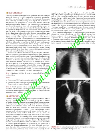
chus, depression of the left mainstem bronchus, and obliteration of the FIGURE 120-5. Ruptured thoracic aorta. A. Chest film showing widened mediastinum.
space between the pulmonary artery and the aorta), frequently the only B. Aortogram from the same patient showing lacerated aorta.
section10.indd 1159 1/20/2015 9:21:09 AM