Page 440 - Clinical Hematology_ Theory _ Procedures ( PDFDrive )
P. 440

424            PART 6  ■  Neoplastic Disorders


                                             Immunohistological Features
                    TABLE        22.4
                                             of Selected T-Cell Neoplasms


                                                            Surface Membrane Markers


                   Type of Neoplasm                         CD3           CD4            CD5            CD8


                   T-PLL                                       +             ±              −            −/+


                   T-LGL                                       +             −              −            +


                   Mycosis fungoides/                          +             +              +            −

                   Sézary syndrome


                   T-PLL, T-cell prolymphocytic leukemia/lymphoma; T-LGL, T-cell large gran-
                   ular lymphocytic leukemia; +, >90%; ±, >50%; −/+, <50%; −, <10%.

                   Source:  Handin  RI,  et  al.  (eds.).  Blood:  Principles  and  Practice  of

                   Hematology, 2nd ed, Philadelphia, PA: Lippincott Williams & Wilkins, 2003.


                                                                                                                           FIGURE  22.5   -cell l  rge gr  nul  r ly  phocytic leuke  i  . Bone
                                                                                                                               rrow core biopsy reve  ls    ly  phoi     ggreg  te with    p  le ger  i-

               cell    orphology  (Fig.  22.5),  i    unophenotyping,    n                                                 n  l center   n   interstiti  l ly  phoi   inf ltr  tion in the     j  cent   re  .

                olecul  r genetics.                                                                                        I    unohistoche  ic  l st  ins i  enti y the ly  phoi     ggreg te   s re  c-

                    Proly  phocytic  leuke  i    is  ch  r  cterize    by      l  rge                                      tive   n   the interstiti  l inf ltr te   s   -LGLs. He   toxylin   n   eosin, 20×

               nu  ber  o   s    ll  ly  phocytes  with  sc  nt  cytopl  s      n                                             gnif c  tion. (Fro  Sun   . Flow Cytometry, Immunohistochemistry,
                                                                                                                           and  Molecular  Genetics   or  Hematologic  Neoplasms,  2n    e  ,
               the  i      ture   e  tures  o   proly  phocytes  in  the  periph-                                          Phil    elphi  , PA: Lippincott Willi    s & Wilkins, 2012.)

               er  l bloo  . Cells o    -PLL o  en h  ve pro  inent nucleolus

                 e  iu   or s    ll in size with convolute   nucle  r outlines.

                 -PLL in peripher  l bloo   exhibits s    ll to   e  iu  , roun                                            Prognosis and Treatm ent

               or irregul  r nuclei rese  bling Séz  ry cells. Pro  inent nucle-                                             -cell PLL is typic  lly   ggressive, but    subset o  p  tients     y

               oli   ppe  r only in    s    ll proportion o  c  ses, but cytopl  s-                                        exhibit   n in  olent ph  se o  v  ri  ble length.   -PLL p  tients

                 ic blebbing is co    on.                                                                                  h  ve      e  i  n surviv  l o  7.5   onths.

                    Leukocytosis  c  n  excee    100  ×  10 /L.  Proly  phocytes                                                First-line ther  py  or p  tients with   -PLL is   le  tuzu    b.
                                                                                 9
                 ust excee   55% o  ly  phoi   cells in the peripher  l bloo  .                                            Eligible p  tients     y be consi  ere    or   llogenic bone     r-

                    In c  ses o    -cell PLL, the i    unophenotypes   re CD2+,                                            row tr  nspl  nt  tion.

               CD3+, CD5+, CD7 +(very strong),   n   CD52 +(very strong).

               Most p  tients   re CD4+. Neg  tive results   re observe    or                                              Sézary Syndrome and Mycosis Fungoides

                     , CD1  ,   n   CD25. Cytogenetic   bnor    lities inclu  e

               Inv  14,  t(14;14),  t(x;14),    n    Iso  8q  co  plex.  Molecul  r                                        T e leuke  ic ph  se o  cut  neous   -cell ly  pho     (C  CL)

               genetics reve  l   ut  tions   CL1, M  CP 1, A  M JAK3,   n                                                 is   ycosis  ungoi  es (MF) (Fig. 22.6). It is the   ost co    on

               S  A  5b.                                                                                                   v  ri  nt o  C  CLs.


                                             Genetic Features and Epstein-

                    TABLE        22.5        Barr Virus Status in Selected

                                             T-Cell Neoplasms


                                                           Genetic

                   Type of Neoplasm                        Abnormality                      EBV Status


                   T-PLL                                   inv 14, trisomy 8q                          −


                   T-LGL                                   None known                                  −


                   Mycosis fungoides/                      None known                                  −

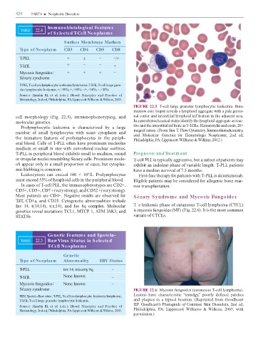
                   Sézary syndrome                                                                                         FIGURE 22.6  Mycosis  ungoi  es (cut  neous   -cell ly  pho    ).


                   EBV Epstein-Barr virus; T-PLL, T-cell prolymphocytic leukemia/lymphoma;                                 Lesions  h  ve  ch  r  cteristic  “s  u  gy,”  poorly    e ine    p  tches
                        ,
                   T-LGL, T-cell large granular lymphocytic leukemia.                                                       n    pl  ques  in      typic  l  loc  tion.  (Reprinte     ro    Goo  he rt
                                                                                                                           HP. Goodheart’s Photoguide o  Common Skin Disorders, 2n   e  ,
                   Source:  Handin  RI,  et  al.  (eds.).  Blood:  Principles  and  Practice  of

                   Hematology, 2nd ed, Philadelphia, PA: Lippincott Williams & Wilkins, 2003.                              Phil    elphi  ,  PA:  Lippincott  Willi    s  &  Wilkins,  2003,  with
                                                                                                                           per  ission.)
   435   436   437   438   439   440   441   442   443   444   445