
Page 375 - fbkCardioDiabetes_2017
P. 375
“Echocardiographic Evaluvation of A Diabetic Patient” 351
Diabetic Cardiomyopathy Hypertension or Valvular heart disease .Three-dimen-
7
sional Echocardiographic stress imaging adds a new
The observation of frequent association of heart dimension.
failure and Diabetes was made by Leyden et al in
1881 . However, it was Rubler in 1972, who introduced
5
the term Diabetic Cardiomyopathy . The minimal cri- Diastolic function in Diabetic
6
teria to diagnose Diabetic Cardiomyopathy include Cardiomyopathy
1. Concentric Left Ventricular Hypertrophy with Abnormal E/A and Deceleration time: Abnormal re-
LV diastolic dysfunction and Interstitial fibrosis laxation characterised by decreased E/A ratio and
2. Eccentric Dilated Cardiomyopathy with re- prolonged Deceleration time and Restrictive pattern
duced left ventricular systolic function with increased E/A ratio and decreased Deceleration
independent of Coronary Artery Disease, Systemic time are well described in early studies 8
Abnormal E/e’ Ratio:
The ratio of early diastolic flow velocity of mitral inflow (E) to early diastolic mitral annular velocity (E/e′) has
been shown to be the most accurate non-invasive marker of elevated LV filling pressure 9,10 . In particular, echo-
cardiographic indices of elevated LV filling pressure are clearly associated with poor cardiac functional and
clinical outcome. E/e′ > 15 is the strongest predictor of cardiac death and readmission for heart failure. (Fig 2)
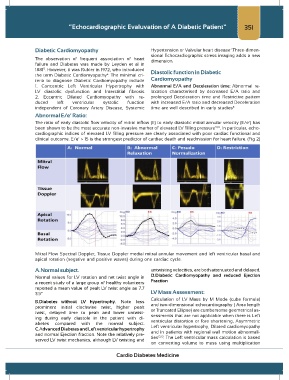
Mitral Flow Spectral Doppler, Tissue Doppler medial mitral annular movement and left ventricular basal and
apical rotation (negative and positive waves) during one cardiac cycle.
A.Normal subject. untwisting velocities, are both attenuated and delayed.
Normal values for LV rotation and net twist angle in D.Diabetic Cardiomyopathy and reduced Ejection
a recent study of a large group of healthy volunteers Fraction
reported a mean value of peak LV twist angle as 7.7
3.5° LV Mass Assessment:
Calculation of LV Mass by M Mode (cube formula)
B.Diabetes without LV hypertrophy. Note less and two-dimensional echocardiography ( Area length
prominent initial clockwise twist, higher peak or Truncated Ellipse) are cumbersome geometrical as-
twist, delayed time to peak and lower untwist- sessments that are not applicable when there is Left
ing during early diastole in the patient with di- ventricular distortion or fore shortening, Asymmetric
abetes compared with the normal subject. Left ventricular hypertrophy, Dilated cardiomyopathy
C.Advanced Diabetes and Left ventricular hypertrophy and in patients with regional wall motion abnormali-
and normal Ejection fraction. Note the relatively pre- ties 12,13 . The Left ventricular mass calculation is based
served LV twist mechanics, although LV twisting and
on converting volume to mass using multiplication
Cardio Diabetes Medicine