
Page 37 - Critical Care Nursing Demystified
P. 37
22 CRITICAL CARE NURSING DeMYSTIFIED
Common carotid artery
Apex of right lung Internal jugular vein
Subclavian
1 artery
Manubrium
Subclavian
vein
Sternum
Fourth rib
Apex of
Xiphoid heart
6
process
Diaphragm
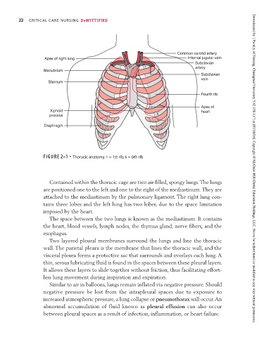
FIGURE 2–1 • Thoracic anatomy. 1 = 1st rib, 6 = 6th rib Downloaded by [ Faculty of Nursing, Chiangmai University 5.62.158.117] at [07/18/16]. Copyright © McGraw-Hill Global Education Holdings, LLC. Not to be redistributed or modified in any way without permission.
Contained within the thoracic cage are two air-filled, spongy lungs. The lungs
are positioned one to the left and one to the right of the mediastinum. They are
attached to the mediastinum by the pulmonary ligament. The right lung con-
tains three lobes and the left lung has two lobes, due to the space limitation
imposed by the heart.
The space between the two lungs is known as the mediastinum. It contains
the heart, blood vessels, lymph nodes, the thymus gland, nerve fibers, and the
esophagus.
Two layered pleural membranes surround the lungs and line the thoracic
wall. The parietal pleura is the membrane that lines the thoracic wall, and the
visceral pleura forms a protective sac that surrounds and overlays each lung. A
thin, serous lubricating fluid is found in the spaces between these pleural layers.
It allows these layers to slide together without friction, thus facilitating effort-
less lung movement during inspiration and expiration.
Similar to air in balloons, lungs remain inflated via negative pressure. Should
negative pressure be lost from the intrapleural spaces due to exposure to
increased atmospheric pressure, a lung collapse or pneumothorax will occur. An
abnormal accumulation of fluid known as pleural effusion can also occur
between pleural spaces as a result of infection, inflammation, or heart failure.