
Page 41 - Critical Care Nursing Demystified
P. 41
26 CRITICAL CARE NURSING DeMYSTIFIED
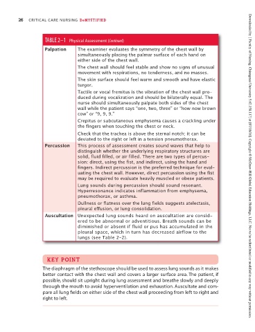
TABLE 2–1 Physical Assessment (Continued)
Palpation The examiner evaluates the symmetry of the chest wall by
simultaneously placing the palmar surface of each hand on
either side of the chest wall.
The chest wall should feel stable and show no signs of unusual
movement with respirations, no tenderness, and no masses.
The skin surface should feel warm and smooth and have elastic
turgor.
Tactile or vocal fremitus is the vibration of the chest wall pro-
duced during vocalization and should be bilaterally equal. The
nurse should simultaneously palpate both sides of the chest
wall while the patient says “one, two, three” or “how now brown
cow” or “9, 9, 9.”
Crepitus or subcutaneous emphysema causes a crackling under
the fingers when touching the chest or neck.
Check that the trachea is above the sternal notch; it can be
deviated to the right or left in a tension pneumothorax.
Percussion This process of assessment creates sound waves that help to
distinguish whether the underlying respiratory structures are
solid, fluid filled, or air filled. There are two types of percus-
sion: direct, using the fist, and indirect, using the hand and
fingers. Indirect percussion is the preferred technique for eval- Downloaded by [ Faculty of Nursing, Chiangmai University 5.62.158.117] at [07/18/16]. Copyright © McGraw-Hill Global Education Holdings, LLC. Not to be redistributed or modified in any way without permission.
uating the chest wall. However, direct percussion using the fist
may be required to evaluate heavily muscled or obese patients.
Lung sounds during percussion should sound resonant.
Hyperresonance indicates inflammation from emphysema,
pneumothorax, or asthma.
Dullness or flatness over the lung fields suggests atelectasis,
pleural effusion, or lung consolidation.
Auscultation Unexpected lung sounds heard on auscultation are consid-
ered to be abnormal or adventitious. Breath sounds can be
diminished or absent if fluid or pus has accumulated in the
pleural space, which in turn has decreased airflow to the
lungs (see Table 2–2).
KEY POINT
The diaphragm of the stethoscope should be used to assess lung sounds as it makes
better contact with the chest wall and covers a larger surface area. The patient, if
possible, should sit upright during lung assessment and breathe slowly and deeply
through the mouth to avoid hyperventilation and exhaustion. Auscultate and com-
pare all lung fields on either side of the chest wall proceeding from left to right and
right to left.