
Page 44 - Critical Care Nursing Demystified
P. 44
Chapter 2 CARE OF THE PATIENT WITH CRITICAL RESPIRATORY NEEDS 29
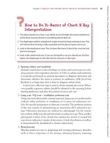
H ? How to Do It—Basics of Chest X-Ray
o
w t
I
—Ba
o
o
s
D
t
Interpretation
Interpretation
y
a
he dark mat
.
1. The dark material on a chest x-ray will be air and the light structures/substances
1
erial on a chest x-r
will be
T
blood
,
,
exudat
e
or somethin
will be fluid, exudate, blood, or something denser than air.
will be fluid
,
i
c
r
l
g
n
agm an
a
op
h
ost
e c
h
re
n
t
d
2.
h
T
2. The diaphragm and the costophrenic angles should be sharp and easy to see. This
iap
d
e
h
will indicate that the lung is fully expanded and the pleural spaces are intact.
wi ll i nd ic at e th at t he l un g is f ul ly e xp an de
3. Look at the mediastinal area. This contains the heart. It should be a normal size
and not enlarged.
4. Look at the abdominal area. If you are facing the x-ray you should see the liver
below the diaphragm on the left and the stomach on the right.
2. Sputum culture and sensitivity
Sputum examination is microbiologic in nature and is necessary in evalu-
ating patients with respiratory disorders. A C&S, or culture and sensitivity, Downloaded by [ Faculty of Nursing, Chiangmai University 5.62.158.117] at [07/18/16]. Copyright © McGraw-Hill Global Education Holdings, LLC. Not to be redistributed or modified in any way without permission.
is routinely performed on sputum specimens to diagnose infections and
determine whether the strain is resistant to antibiotics. AFB (Acid Fast
Bacillus) is a Gram stain that is done to diagnose tuberculosis.
A specimen collection trap is used to obtain sputum specimens. When-
ever possible a sputum culture should be obtained in the morning, before
starting antibiotics and after the patient receives oral care.
3. Lung scan –VQ scan – ventilation perfusion scan
Using injected radionuclide contrast material, lung scans are performed to
evaluate either perfusion or ventilation, or to assess for pulmonary em-
boli. No specific preparation or aftercare is needed. The perfusion portion
of the test consists of administering an intravenous (IV) radioactive iso-
tope. Pulmonary structures are outlined in a photograph. For the ventila-
tion portion of the test, the patient inhales a radioactive gas. Then another
photograph is taken of the alveoli that uptakes the alveoli. A normal VQ
scan shows radioactive uptake of structures. A lack of perfusion or airflow
is demonstrated by diminished or absent radioactivity.
4. Bronchoscopy
This has numerous uses in diagnosing and treating pulmonary disorders
such as direct inspection of the airways, obtaining biopsies, removing