
Page 287 - Color_Atlas_of_Physiology_5th_Ed._-_A._Despopoulos_2003
P. 287
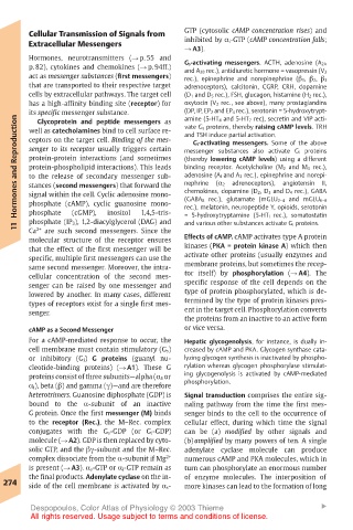
Cellular Transmission of Signals from  GTP (cytosolic cAMP concentration rises) and
       Extracellular Messengers        inhibited by α i-GTP (cAMP concentration falls;
                                       ! A3).
       Hormones, neurotransmitters (! p. 55 and
       p. 82), cytokines and chemokines (! p. 94ff.)  G s-activating messengers. ACTH, adenosine (A 2A
                                       and A 2B rec.), antidiuretic hormone = vasopressin (V 2
       act as messenger substances (first messengers)  rec.), epinephrine and norepinephrine (" 1, " 2, " 3
       that are transported to their respective target  adrenoceptors), calcitonin, CGRP, CRH, dopamine
       cells by extracellular pathways. The target cell  (D 1 and D 5 rec.), FSH, glucagon, histamine (H 2 rec.),
       has a high-affinity binding site (receptor) for  oxytocin (V 2 rec., see above), many prostaglandins
       its specific messenger substance.  (DP, IP, EP 2 and EP 4 rec.), serotonin = 5-hydroxytrypt-
                                       amine (5-HT 4 and 5-HT 7 rec), secretin and VIP acti-
    Hormones and Reproduction  protein-protein interactions (and sometimes  messenger substances also activate G i proteins
         Glycoprotein and peptide messengers as
                                       vate G s proteins, thereby raising cAMP levels. TRH
       well as catecholamines bind to cell surface re-
                                       and TSH induce partial activation.
       ceptors on the target cell. Binding of the mes-
                                        G i-activating messengers. Some of the above
       senger to its receptor usually triggers certain
                                       (thereby lowering cAMP levels) using a different
       protein-phospholipid interactions). This leads
                                       binding receptor. Acetylcholine (M 2 and M 4 rec.),
                                       adenosine (A l and A 3 rec.), epinephrine and norepi-
       to the release of secondary messenger sub-
                                       nephrine
                                                           angiotensin
                                                                   II,
                                                adrenoceptors),
       stances (second messengers) that forward the
                                             (α 2
                                       chemokines, dopamine (D 2, D 3 and D 4 rec.), GABA
       signal within the cell. Cyclic adenosine mono-
                                       rec.), melatonin, neuropeptide Y, opioids, serotonin
       phosphate
                       inositol
                              1,4,5-tris-
                (cGMP),
                                       = 5-hydroxytryptamine (5-HT l rec.), somatostatin
       phosphate (IP 3), 1,2-diacylglycerol (DAG) and
                                       and various other substances activate G i proteins.
    11  phosphate (cAMP), cyclic guanosine mono-  (GABA B rec.), glutamate (mGLU 2–4 and mGLU 6–8
       Ca
          are such second messengers. Since the
        2+
       molecular structure of the receptor ensures  Effects of cAMP. cAMP activates type A protein
       that the effect of the first messenger will be  kinases (PKA = protein kinase A) which then
       specific, multiple first messengers can use the  activate other proteins (usually enzymes and
       same second messenger. Moreover, the intra-  membrane proteins, but sometimes the recep-
       cellular concentration of the second mes-  tor itself) by phosphorylation (! A4). The
       senger can be raised by one messenger and  specific response of the cell depends on the
       lowered by another. In many cases, different  type of protein phosphorylated, which is de-
       types of receptors exist for a single first mes-  termined by the type of protein kinases pres-
       senger.                         ent in the target cell. Phosphorylation converts
                                       the proteins from an inactive to an active form
       cAMP as a Second Messenger      or vice versa.
       For a cAMP-mediated response to occur, the  Hepatic glycogenolysis, for instance, is dually in-
       cell membrane must contain stimulatory (G s)  creased by cAMP and PKA. Glycogen synthase cata-
       or inhibitory (G i) G proteins (guanyl nu-  lyzing glycogen synthesis is inactivated by phospho-
       cleotide-binding proteins) (! A1). These G  rylation whereas glycogen phosphorylase stimulat-
       proteins consist of three subunits—alpha (α S or  ing glycogenolysis is activated by cAMP-mediated
       α i), beta (") and gamma (γ)—and are therefore  phosphorylation.
       heterotrimers. Guanosine diphosphate (GDP) is  Signal transduction comprises the entire sig-
       bound to the α-subunit of an inactive  naling pathway from the time the first mes-
       G protein. Once the first messenger (M) binds  senger binds to the cell to the occurrence of
       to the receptor (Rec.), the M–Rec. complex  cellular effect, during which time the signal
       conjugates with the G s-GDP (or G i-GDP)  can be (a) modified by other signals and
       molecule (! A2). GDP is then replaced by cyto-  (b) amplified by many powers of ten. A single
       solic GTP, and the "γ-subunit and the M–Rec.  adenylate cyclase molecule can produce
       complex dissociate from the α-subunit if Mg 2+  numerous cAMP and PKA molecules, which in
       is present (! A3). α s-GTP or α i-GTP remain as  turn can phosphorylate an enormous number
       the final products. Adenylate cyclase on the in-  of enzyme molecules. The interposition of
  274  side of the cell membrane is activated by α s-  more kinases can lead to the formation of long
       Despopoulos, Color Atlas of Physiology © 2003 Thieme        !
       All rights reserved. Usage subject to terms and conditions of license.