
Page 346 - Color_Atlas_of_Physiology_5th_Ed._-_A._Despopoulos_2003
P. 346
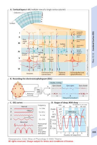
A.  Cortical layers I–VI (multiple view of a single-cortex column)
           Columns              I
                                II
                                III
                                IV
                                V
                               VI
        Surface
                  1           2           3          4
               I                                          Unspecific
                                                          thalamo-
                                                          cortical
               II                                         fibers
                  Pyra-                                   Commissural
                  midal                                   fibers       Cerebral Cortex EEG
               III  cell                      Stellate
                              Axon              cell      Association
                                                          fibers
                                                          Specific
              IV             Stellate                     thalamo-
                 Principal                                cortical
                 dendrite    cell                         fibers
                        Pyramidal  Associa-
                                      Com-
               V        cell    tion  missural                         Plate 12.12
                                fibers  fibers                    (After  Szentágothai and Birbaumer/Schmidt)
                             Axon
                             colla-
              VI             teral
                                Corticocortical  Thalamic  Corticobulbar and
                     Neurons
                                connections  afferents  spinal efferents
         B.  Recording the electroencephalogram (EEG)
                                      Awake, resting
               1                        Eyes closed  Eyes open  Eyes closed
                                    1
                            EEG
                            unit
                     Reference
                     electrode      7
               7                           α         β        α
                                               “Desynchronization”
         C.  EEG curves                D.  Stages of sleep, REM sleep
                             Frequency       EEG       (See also text on next page)
                 Normal                         10  20   30  40  min
         α                    8–13Hz   Awake  α/β
         β           100µV    14–30Hz   A    α/ϑ    REM sleep
         ϑ                     4–7Hz    B(1)  ϑ
                      1s               REM    ϑ
         δ                    0.5–3Hz   C(2)  ϑ
                Abnormal
                             Paroxysmal  Stage of sleep  D(3)  δ
                             spikes
                             Paroxysmal  E(4)  δ                  c)  ´
                             waves
                             3Hz spikes                           (After Iovanovi`  333
                             and waves     0  1  2  3  4  5  6
                                                Duration of sleep (hrs)
       Despopoulos, Color Atlas of Physiology © 2003 Thieme
       All rights reserved. Usage subject to terms and conditions of license.