
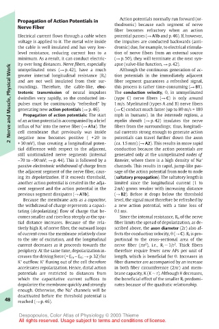
Page 61 - Color_Atlas_of_Physiology_5th_Ed._-_A._Despopoulos_2003
P. 61
Propagation of Action Potentials in  Action potentials normally run forward (or-
       Nerve Fiber                     thodromic) because each segment of nerve
                                       fiber becomes refractory when an action
       Electrical current flows through a cable when  potential passes (! A1b and p. 46). If, however,
       voltage is applied to it. The metal wire inside  the impulses are conducted backwards (anti-
       the cable is well insulated and has very low-  dromic) due, for example, to electrical stimula-
       level resistance, reducing current loss to a  tion of nerve fibers from an external source
       minimum. As a result, it can conduct electric-  (! p. 50), they will terminate at the next syn-
       ity over long distances. Nerve fibers, especially  apse (valve-like function, ! p. 42).
    Nerve and Muscle, Physical Work  roundings. Therefore, the cable-like, elec-  this process is rather time-consuming (! B1).
       unmyelinated ones (! p. 42), have a much
                                        Although the continuous generation of ac-
                                       tion potentials in the immediately adjacent
       greater internal longitudinal resistance (R i)
       and are not well insulated from their sur-
                                       fiber segment guarantees a refreshed signal,
                                       The conduction velocity, θ, in unmyelinated
       trotonic transmission of neural impulses
                                       (type C) nerve fibers (! C) is only around
       dwindles very rapidly, so the conducted im-
                                       1 m/s. Myelinated (types A and B) nerve fibers
       pulses must be continuously “refreshed” by
                                       (! C) conduct much faster (up to 80 m/s = 180
       generating new action potentials (! p. 46).
                                       mph in humans). In the internode regions, a
         Propagation of action potentials: The start
                                       myelin sheath (! p. 42) insulates the nerve
       of an action potential is accompanied by a brief
              +
       cell membrane that previously was inside
                                       nal currents strong enough to generate action
                                       potentials can travel further down the axon
       negative now becomes positive ( + 20 to
    2  influx of Na into the nerve fiber (! A1a). The  fibers from the surroundings; thus, longitudi-
       + 30 mV), thus creating a longitudinal poten-
                                       (ca. 1.5 mm) (! A2). This results in more rapid
       tial difference with respect to the adjacent,  conduction because the action potentials are
       still unstimulated nerve segments (internal  generated only at the unmyelinated nodes of
       –70 to –90 mV; ! p. 44). This is followed by a  Ranvier, where there is a high density of Na +
       passive electrotonic withdrawal of charge from  channels. This results in rapid, jump-like pas-
       the adjacent segment of the nerve fiber, caus-  sage of the action potential from node to node
       ing its depolarization. If it exceeds threshold,  (saltatory propagation). The saltatory length is
       another action potential is created in the adja-  limited since the longitudinal current (1 to
       cent segment and the action potential in the  2 nA) grows weaker with increasing distance
       previous segment dissipates (! A1b).  (! B2). Before it drops below the threshold
         Because the membrane acts as a capacitor,  level, the signal must therefore be refreshed by
       the withdrawal of charge represents a capaci-  a new action potential, with a time loss of
       tating (depolarizing) flow of charge that be-  0.1 ms.
       comes smaller and rises less steeply as the spa-  Since the internal resistance, R i, of the nerve
       tial distance increases. Because of the rela-  fiber limits the spread of depolarization, as de-
       tively high R i of nerve fiber, the outward loops  scribed above, the axon diameter (2r) also af-
       of current cross the membrane relatively close  fects the conduction velocity, θ (! C). R i is pro-
       to the site of excitation, and the longitudinal  portional to the cross-sectional area of the
                                                 2
                                                          2
       current decreases as it proceeds towards the  nerve fiber (πr ), i.e., R i ! 1/r . Thick fibers
       periphery. At the same time, depolarization in-  therefore require fewer new APs per unit of
       creases the driving force (= E m – E K; ! p. 32) for  length, which is beneficial for θ. Increases in
       K outflow. K fluxing out of the cell therefore  fiber diameter are accompanied by an increase
               +
        +
       accelerates repolarization. Hence, distal action  in both fiber circumference (2πr) and mem-
       potentials are restricted to distances from  brane capacity, K (K ! r). Although θ decreases,
       which the capacitative current suffices to  the beneficial effect of the smaller R i predomi-
       depolarize the membrane quickly and strongly  nates because of the quadratic relationship.
                        +
       enough. Otherwise, the Na channels will be
       deactivated before the threshold potential is
   48  reached (! p. 46).
       Despopoulos, Color Atlas of Physiology © 2003 Thieme
       All rights reserved. Usage subject to terms and conditions of license.