
Page 1015 - Hall et al (2015) Principles of Critical Care-McGraw-Hill
P. 1015
746 PART 5: Infectious Disorders
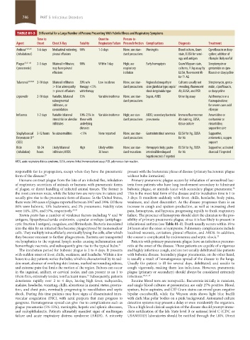
TABLE 81-3 Differential for a Large Number of Persons Presenting With Febrile Illness and Respiratory Symptoms
Time to Onset to- Person-to-
Agent Onset Chest X-Ray Fatality Respiratory Failure Person Infection Complications Diagnosis Treatment
Anthrax 14,25,26 1-6 days Mediastinal widening; 90% 1-3 days None; use stan- Meningitis Blood culture, Gram Ciprofloxacin or doxy-
(inhalational) pleural effusions dard precautions stain, ELISA for serol- cycline; addition of
ogy and antigen rifampin likely useful
Plague 35,42–44 2-3 days Bilateral infiltrates; 90% Within 1 day High; use Early hemoptysis Gram/Wayson stain, Streptomycin or
(pneumonic) may have pleural respiratory cultures, Fl Ag assay by gentamicin or cipro-
effusions isolation ELISA, fluorescent Ab floxacin or doxycycline
for F1 AG
Tularemia 53,55,58 2-10 days Bilateral infiltrates 30% w/o Low incidence None; use stan- Regional adenopathy in Cultures usually not Streptomycin, genta-
> hilar adenopathy therapy; <5% dard precautions ulcer glandular type; sepsis/ revealing; fluorescent micin, ciprofloxacin,
> pleural effusions with therapy shock in typhoidal type Ab, ELISA, and PCR or doxycycline
Legionella 2-10 days Variable, bilateral 15% Variable incidence None; use stan- Sepsis, ARDS Urine Ag assay Azithromycin or a
subsegmental dard precautions fluoroquinolone;
infiltrates, or for severe cases add
consolidation rifampin
Influenza 1-2 days Variable bilateral 10%-25% in Variable incidence High; use stan- ARDS; secondary bacterial Immunofluorescence Amantidine or
interstitial or alveolar those with dard precautions pneumonia Ab staining, ELISA, oseltamivir or
infiltrates underlying tissue culture rimantidine;
diseases supportive care
Staphylococcal 3-12 hours No abnormalities <1% None reported None; use stan- Gastrointestinal anorexia ELISA for Ag, ELISA Supportive,
Enterotoxin B dard precautions for Ab antiemetics, oxygen
24
(SEB) support
Ricin 18-24 Likely bilateral High Likely within None; use stan- Hemoptysis likely; gastro- ELISA for Ag, ELISA Supportive; activated
( inhalation) hours infiltrates/ARDS 30 hours dard recautions intestinal bleeding and for Ab charcoal if ingested
hepatic necrosis if ingested
ARDS, acute respiratory distress syndrome; ELISA, enzyme-linked immunosorbent assay; PCR, polymerase chain reaction.
responsible for its propagation, except when they have the pneumonic present with the bacteremic phase of disease ( primary bacteremic plague
form of the disease. 29 without bubo formation).
Humans contract plague from the bite of an infected flea, inhalation Primary pneumonic plague occurs by inhalation of aerosolized bac-
of respiratory secretions of animals or humans with pneumonic forms teria from patients who have lung involvement secondary to fulminant
of plague, or direct handling of infected animal tissues. The former is bubonic plague, or animals (cats) with secondary plague pneumonia.
32
the most common route, while the latter two are very rare in nature and This is the most fatal form of the disease and its incubation time is 1 to
usually give rise to the pneumonic form of disease. In the United States, 3 days. It manifests suddenly with fever, chills, headache, body pains,
there were 390 cases of plague reported between 1947 and 1996. Of these weakness, and chest discomfort. As the disease progresses there is an
84% were bubonic, 13% bacteremic, and 2% pneumonic. Fatality rates increase in cough and sputum production, as well as increasing chest
were 14%, 22%, and 57%, respectively. 29,30 pain, hemoptysis, and hypoxia, progressing rapidly to frank respiratory
Yesinia pestis has a number of virulence factors including V and W failure. The presence of hemoptysis should alert the clinician to the pos-
antigens, lipopolysaccharide endotoxin, capsular envelope (antiphago- sibility of primary pneumonic plague, since it is less likely to present in
cytic fraction I antigen), coagulase, and fibrinolysin. Bacteria inoculated inhalational anthrax (see Table 81-3). Death usually occurs within 18 to
into the skin by an infected flea become phagocytosed by mononuclear 24 hours after the onset of symptoms. Pulmonary complications include
cells. They multiply intracellularly, eventually lysing the cells, after which localized necrosis, cavitation, pleural effusion, and ARDS. In addition,
they become resistant to further phagocytosis. Bacteria are transported the course is complicated by endotoxemia and septic shock. 33
via lymphatics to the regional lymph nodes causing inflammation and Patients with primary pneumonic plague have an infectious pneumo-
hemorrhagic necrosis, and subsequently give rise to the typical bubo. 31 nitis at the onset of the disease. These patients are capable of a vigorous
The incubation period for bubonic plague is 2 to 8 days. It presents and highly infectious cough, and are not usually debilitated like patients
with sudden onset of fever, chills, weakness, and headache. Within a few with bubonic disease. Secondary plague pneumonia, on the other hand,
hours to a day patients notice the bubo, which is characterized by its sud- is usually a result of hematogenous spread of the disease to the lungs.
den onset, absence of overlying skin lesions, marked surrounding edema, Usually the patient is ill for several days, debilitated, and unable to
and extreme pain that limits the motion of the region. Buboes can occur cough vigorously, making them less infectious. However, pneumonic
in the inguinal, axillary, or cervical nodes, and can present as an 1 to plague (primary or secondary) should always be considered extremely
10 cm firm, extremely tender, nonfluctuant mass. Subsequently, patients infectious. 29,31,33
31
deteriorate rapidly over 2 to 4 days, having high fever, tachycardia, Routine blood tests are nonspecific. Bacteremia initially is transient,
malaise, headache, vomiting, chills, alterations in mental status, prostra- and single blood cultures at presentation are only 27% positive. Blood,
tion, and chest pain, eventually progressing to vasodilation and septic sputum, bubo aspirates, and CSF Gram stains can reveal gram-negative
shock. During this time patients may have signs of disseminated intra- bipolar coccobacilli, while the Wayson stain shows light blue bacilli
vascular coagulation (DIC), with acral purpura that may progress to with dark blue polar bodies on a pink background. Automated culture
gangrene. Hematogenous spread can give rise to complications such as detection systems may present a delay or even misidentify the organism.
plague pneumonia (5%-15%), meningitis, hepatic and splenic abscesses, Thus a high level clinical suspicion of the disease should prompt imme-
and endophthalmitis. Patients ultimately manifest signs of multiorgan diate notification of the lab. State level B or national level C (CDC or
failure and acute respiratory distress syndrome (ARDS). A minority USAMRIID) laboratories should be notified through the LRN. Direct
section05_c74-81.indd 746 1/23/2015 12:37:44 PM